Chapter: Clinical Dermatology: Diagnosis of skin disorders
Special tools and techniques - Diagnosis of skin disorders
Special
tools and techniques
A magnifying lens is a helpful aid to diagnosis because subtle changes in the skin become more apparent when enlarged. One attached to spectacles will leave your hand free.
A
Wood’s
light, emitting long wavelength ultraviolet radiation, will help
with the examination of some skin conditions. Fluorescence is seen in some
fungal infections , erythrasma and
pseudomonas infections. Some subtle disorders of pigmentation can be seen more
clearly under Wood’s light, e.g. the pale patches of tuberous sclerosis,
low-grade vitiligo and pityriasis versicolor, and the darker café-au-lait
patches of neurofibromatosis. The urine inhepatic cutaneous
porphyria often fluoresces coral pink, even without solvent extraction of the
porphyrins (see Fig. 19.10).
Diascopy is
the name given to the technique in whicha glass slide or clear plastic spoon is
used to blanch vascular lesions and so to unmask their underlying colour.
Photography,
conventional or digital, helps to recordthe baseline appearance of a lesion or
rash,
so that change can be assessed objectively at later visits. Small changes in
pigmented lesions can be detected by ana-lysing sequential digital images
stored in computerized systems.
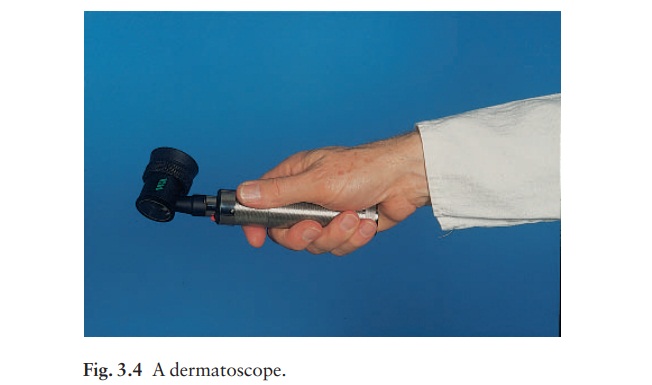
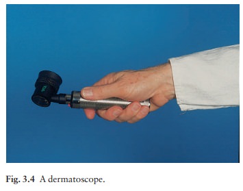
Dermatoscopy (epiluminescence microscopy, skin surface microscopy)
This
non-invasive technique for diagnosing pigmented lesions in vivo
has come of age in the last few years. It is particularly useful in the
diagnosis of malignant melanomas. The lesion is covered with mineral
oil, alcohol or water and then illuminated and observed at
The fluid eliminates
surface reflection and makes the horny layer translucent so that pigmented
structures in the epidermis and superficial dermis and the superficial vascular
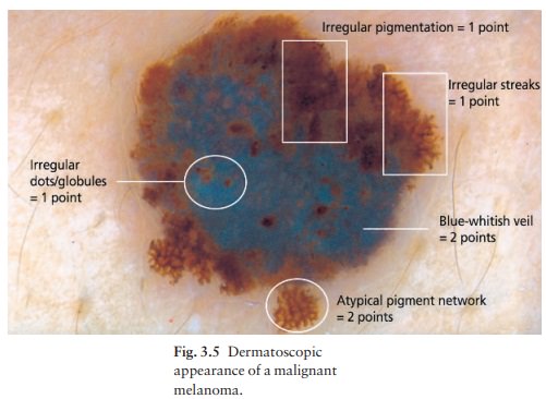
plexus can be assessed. The dermatoscopic appearance of many pigmented lesions,
including seborrhoeic warts, haemangiomas, basal cell carcinomas and most naevi
and malignant melanomas is characteristic (Fig. 3.5). Images can be recorded by
conventional or digital photography and sequential changes assessed. With
formal training and practice, the use of dermatoscopy improves
the accur-acy with which pigmented lesions are diagnosed.
A
dermatoscope can also be used to identify scabies mites in their burrows.
Related Topics