Chapter: Essentials of Anatomy and Physiology: Muscular System
Smooth Muscle and Cardiac Muscle
SMOOTH MUSCLE AND CARDIAC MUSCLE
Smooth muscle cells are small and spindle-shaped, usually with one nucleus per cell (table 7.2). They contain less actin and myosin than do skeletal muscle cells, and the myofilaments are not organized into sarcomeres. As a result, smooth muscle cells are not striated. Smooth muscle cells contract more slowly than skeletal muscle cells when stimulated by neurotransmitters from the nervous system and do not develop an oxygen deficit. The rest-ing membrane potential of some smooth muscle cells fluctuates between slow depolarization and repolarization phases. As a result, smooth muscle cells can periodically and spontaneously generate action potentials that cause the smooth muscle cells to contract. The resulting periodic spontaneous contraction of smooth muscle is calledautorhythmicity. Smooth muscle is under involuntary control, whereas skeletal muscle is under voluntary motor control. Some hormones, such as those that regulate the digestive system, can stimulate smooth muscle to contract.
Smooth muscle cells are organized to form layers. Most of those cells have gap junctions, specialized cell-to-cell contacts , that allow action potentials to spread to all the smooth muscle cells in a tissue. Thus, all the smooth muscle cells tend to function as a unit and contract at the same time.
Cardiac muscle shares some characteristics with both smooth and skeletal muscle (table 7.2). Cardiac muscle cells are long, stri-ated, and branching, with usually only one nucleus per cell. The actin and myosin myofilaments are organized into sarcomeres, but the distribution of myofilaments is not as uniform as in skeletal muscle. As a result, cardiac muscle cells are striated, but not as distinctly striated as skeletal muscle. When stimulated by neu-rotransmitters, the rate of cardiac muscle contraction is between
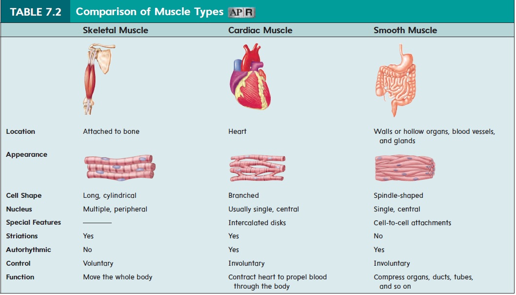
those of smooth and skeletal muscle. Cardiac muscle contraction is autorhythmic. Cardiac muscle exhibits limited anaerobic respi-ration. Instead, it continues to contract at a level that can be sus-tained by aerobic respiration and consequently does not fatigue.
Cardiac muscle cells are connected to one another by inter-calated (in-ter′ka-lā-ted) disks. Intercalated disks are specializedstructures that include tight junctions and gap junctions and that facilitate action potential conduction between the cells. This cell-to-cell connection allows cardiac muscle cells to function as a unit. As a result, an action potential in one cardiac muscle cell can stimulate action potentials in adjacent cells, causing all to contract together. As with smooth muscle, cardiac muscle is under involun-tary control and is influenced by hormones, such as epinephrine.
Related Topics