Chapter: Medicine and surgery: Cardiovascular system
Signs of Oedema
Signs
Oedema
Oedema is defined as an abnormal accumulation of fluid within the
interstitial spaces. A number of mechanisms are thought to be involved in the
development of oedema. Normally tissue fluid is formed by a balance of
hydrostatic and osmotic pressure.
Hydrostatic pressure is the pressure within the blood vessel (high in
arteries, low in veins). Oncotic pressure is produced by the large molecules
within the blood (albumin, haemoglobin) and draws water osmotically back into
the vessel. The hydrostatic pressure is high at the arterial end of a capillary
bed hence fluid is forced out of the vasculature (see Fig. 2.1).
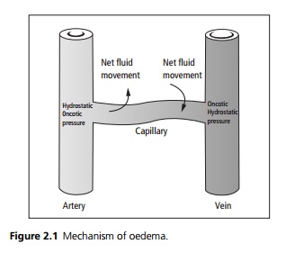
The colloid osmotic pressure then draws fluid back in at the venous end of the capillary bed as the hydrostatic pressure of the venules is low. Any remaining interstitial fluid is then
returned to the circulation via the lymphatic system.
Mechanisms of cardiovascular oedema include the following:
·
Raised venous pressure raising
the hydrostatic pressure at the venous end of the capillary bed (right
ventricular failure, pericardial constriction, vena caval obstruction).
·
Salt and water retention
occurring in heart failure, which increases the circulating blood volume with
pooling on the venous side again raising the hydrostatic pressure.
·
The liver congestion that occurs
in right-sided heart failure may reduce hepatic function, including albumin
production. Albumin is the major factor responsible for the generation of the
colloid osmotic pressure that returns the tissue fluid to the vasculature. A
drop in albumin therefore results in an accumulation of oedema.
Oedema is described as pitting (an indentation or pit is left after
pressing with a thumb for several seconds) or nonpitting. Cardiac oedema is
pitting unless long standing when secondary changes in the lymphatics may cause
a nonpitting oedema. Distribution is dependent on the patient. Patients who are
confined to bed develop oedema around the sacral area rather than the classical
ankle and lower leg distribution. Pleural effusions and ascites may develop in
severe failure.
Related Topics