Chapter: Human Nervous System and Sensory Organs : Brain Stem and Cranial Nerves
Red Nucleus and Substantia Nigra
Red Nucleus and Substantia Nigra
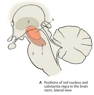
Lateral View of the Brain Stem (A)
The two large nuclei extend far toward the diencephalon. The substantia nigra (AB1) reaches from the oral part of the pons (A2) to the pallidum(AB3) in the diencephalon. Both nuclei are important relay stations of the extrapyramidal system.
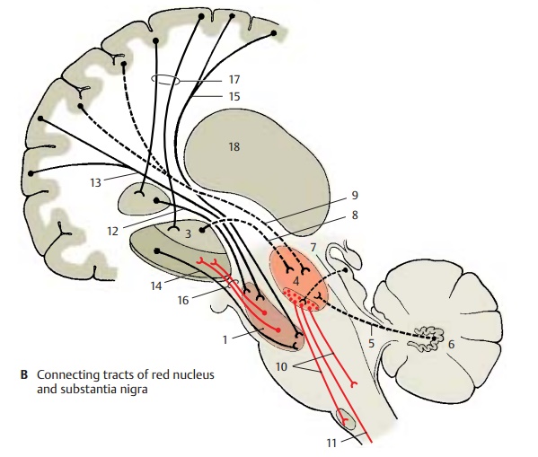
Red Nucleus (A, B)
The nucleus (AB4) appears reddish in a fresh brain section (high iron content, p. 148, A). It consists of the parvocellular neorubrum and the magnocellular paleoru-brum situated ventrocaudally.
Afferent connections
The dentatorubral fasciculus (B5) of the dentate nucleus (B6) of the cerebellumruns in the superior cerebellar peduncle and terminates in the contralateral red nucleus.

! The tectorubral tract (B7) of the superior colliculus terminates in the ipsilateral and contralateral paleorubrum.
! The pallidorubral tract (B8) consists of pallidotegmental bundles from the inner segment of the pallidum.
! The corticorubral tract (B9) from the fron-tal and precentral cortex terminates in the ipsilateral red nucleus.
Efferent connections
The rubroreticular and rubro-olivaryfibers (B10) run in the central tegmental tract and terminate primarilyin the olive (neuronal circuit: dentate nu-cleus – red nucleus – olive – cerebellum).
! The rubrospinal tract (B11) (poorly developed in humans) crosses in Forel’s tegmental decussation and terminates in the cervical spinal cord.
Functional significance.The red nucleus isa relay and control station for cerebellar, pallidal, and corticomotor impulses that are important for muscle tone, posture, and loco-motion. Injury to this nucleus causes passivetremor (shaking), changes in muscle tone, and choreic-athetoid hyperactivity.
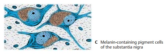
Substantia Nigra (A – C)
This consists of the dark compact part (nerve cells with black melanin pigment) (C) and the reticular part (of reddish color and rich in iron). The tracts of the substantia nigra form only loose pathways of fine fibers rather than compact bundles.
Afferent connections terminating in the anterior part
! Fibers of the caudate nucleus, strionigralfasciculus (B12)
! Fibers of the frontal cortex (areas 9 to 12), corticonigral fibers (B13)
Afferent connections terminating in the caudal part
Fibers of the putamen (B14)
Fibers of the precentral cortex (areas 4 and 6) (B15)
Efferent connections
Nigrostriatal fibers (B16), running from thecompact part to the striatum
Fibers of the reticular part, running to the thalamus
The majority of efferent fibers ascend to the striatum, to which the substantia nigra is closely connected functionally by the nigro-striatal system. In the axons of thedopaminer-gic nigral neurons (compact part), dopamineis transported to the striatum, where it is re-leased by the axon terminals. There is a topographical relationship between the substantia nigra and the striatum (caudate nucleus and putamen); cranial and caudal segments of the substantia nigra are con-nected with the corresponding segments of caudate nucleus and putamen. Caudate nu-cleus and putamen are under the control of massive input from totally different neocor-tical zones (B17).
Functional significance.The substantianigra is of special importance for the control of involuntary coordinated movement and the rapid onset of movement (starter func-tion). Injury causes muscle stiffness, passivetremor, and loss of coordinated movement and facial expression (masklike expression ofthe face).
B18 Posterior thalamus.
Related Topics