Chapter: Medical Physiology: Rhythmical Excitation of the Heart
Rapid Transmission in the Ventricular Purkinje System
Rapid Transmission in the Ventricular Purkinje System
Special Purkinje fibers lead from the A-V node through the A-V bundle into the ventricles. Except for the initial portion of these fibers where they penetrate the A-V fibrous barrier, they have functional charac-teristics that are quite the opposite of those of the A-V nodal fibers. They are very large fibers, even larger than the normal ventricular muscle fibers, and they transmit action potentials at a velocity of 1.5 to 4.0 m/sec, a velocity about 6 times that in the usual ventricular muscle and 150 times that in some of the A-V nodal fibers. This allows almost instantaneous transmission of the cardiac impulse throughout the entire remainder of the ventricular muscle.
The rapid transmission of action potentials by Purk-inje fibers is believed to be caused by a very high level of permeability of the gap junctions at the intercalated discs between the successive cells that make up the Purkinje fibers. Therefore, ions are transmitted easily from one cell to the next, thus enhancing the velocity of transmission. The Purkinje fibers also have very few myofibrils, which means that they contract little or not at all during the course of impulse transmission.
One-Way Conduction Through the A-V Bundle. A specialcharacteristic of the A-V bundle is the inability, except in abnormal states, of action potentials to travel back-ward from the ventricles to the atria. This prevents re-entry of cardiac impulses by this route from the ventricles to the atria, allowing only forward conduc-tion from the atria to the ventricles.
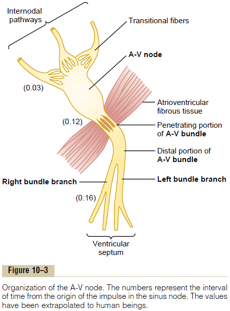
Furthermore, it should be recalled that everywhere, except at the A-V bundle, the atrial muscle is sepa-rated from the ventricular muscle by a continuous fibrous barrier, a portion of which is shown in Figure 10–3. This barrier normally acts as an insulator to prevent passage of the cardiac impulse between atrial and ventricular muscle through any other route besides forward conduction through the A-V bundle itself. (In rare instances, an abnormal muscle bridge does penetrate the fibrous barrier elsewhere besides at the A-V bundle. Under such conditions, the cardiac impulse can re-enter the atria from the ventricles and cause a serious cardiac arrhythmia.)
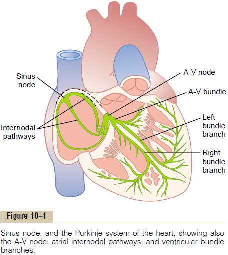
Distribution of the Purkinje Fibers in the Ventricles—The Left and Right Bundle Branches. After penetrating the fibroustissue between the atrial and ventricular muscle, the distal portion of the A-V bundle passes downward in the ventricular septum for 5 to 15 millimeters toward the apex of the heart, as shown in Figures 10–1 and 10–3. Then the bundle divides into left and right bundlebranches that lie beneath the endocardium on the tworespective sides of the ventricular septum. Each branch spreads downward toward the apex of the ven-tricle, progressively dividing into smaller branches. These branches in turn course sidewise around each ventricular chamber and back toward the base of the heart. The ends of the Purkinje fibers penetrate about one third the way into the muscle mass and finally become continuous with the cardiac muscle fibers.
From the time the cardiac impulse enters the bundle branches in the ventricular septum until it reaches the terminations of the Purkinje fibers, the total elapsed time averages only 0.03 second. Therefore, once the cardiac impulse enters the ventricular Purkinje con-ductive system, it spreads almost immediately to the entire ventricular muscle mass.
Related Topics