Chapter: Human Neuroanatomy(Fundamental and Clinical): Autonomic Nervous System
Preganglionic and Postganglionic Neurons
PREGANGLIONIC AND POSTGANGLIONIC NEURONS
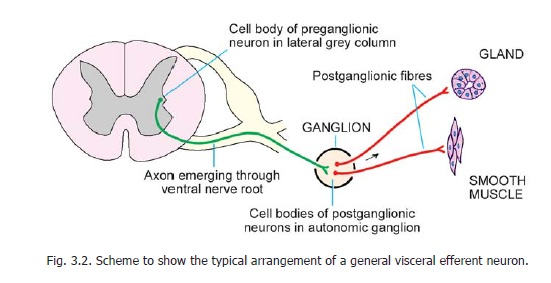
The autonomic pathway, for innervation of smooth muscle or gland always consists of two neurons that synapse in a ganglion (Fig. 3.2). The first neuron carries the nerve impulse from the CNS to the ganglion and is called the preganglionic neuron. The second neuron carries impulses from the ganglion to smooth muscle or gland and is called the postganglionic neuron.
Sympathetic Preganglionic Neurons
The cell bodies of sympathetic preganglionic neurons are located in the intermediolateral grey column of the spinal cord in the thoracic and upper two (or three) lumbar segments (Fig. 19.3). Fibres arising from these neurons constitute the thoracolumbar outflow. Their axons leave the spinal cord through anterior nerve roots to reach the spinal nerves of the segments concerned. After a very short course in the ventral primary rami these fibres enter the white rami communicantes to reach the sympathetic trunk (Fig. 19.1).
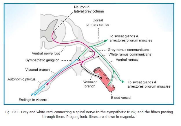
On reaching the sympathetic trunk these fibres behave in one of the following ways (Figs. 19.1, 19.4).
a. They may terminate in relation to cells of the sympathetic ganglion at the level concerned.

b. They may travel up or down the sympathetic trunk to terminate in ganglia at a higher or lower level.
c. They may leave the sympathetic trunk through one of its branches to terminate in a peripherally situated ganglion.

Sympathetic Postganglionic Neurons
These neurons are located primarily in ganglia on the sympathetic trunks. Some are located in peripheral autonomic plexuses (Figs 19.1, 19.5). Axons arising from sympathetic postganglionic neurons behave in one of the following ways.
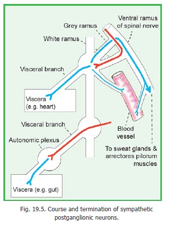
1. The axons may pass through a grey ramus communicans to reach a spinal nerve. They then pass through the spinal nerve and its branches to innervate sweat glands and arrectores pilorum muscles of the skin in the region to which the spinal nerve is distributed.
2. The axons may reach a cranial nerve through a communicating branch and may be distributed through it as in the case of a spinal nerve.
3. The axons may pass into a vascular branch and may be distributed to branches of the vessel. Some fibres from these plexuses may pass to other structures in the neighbourhood of the vessel.
4. We have already seen that some axons, meant for innervation of blood vessels, travel for part of their course in spinal nerves or their branches, and reach the vessels through vascular branches arising from these nerves. Many blood vessels in the peripheral parts of the limbs are innervated in this way.
5. The axons of postganglionic neurons arising in sympathetic ganglia may travel through visceral branches and through autonomic plexuses to reach some viscera (e.g., the heart).
6. The axons of postganglionic neurons located in peripheral autonomic plexuses innervate neighbouring viscera. These fibres often travel to the viscera in plexuses along blood vessels. For example, fibres for the gut travel along plexuses surrounding the branches of the coeliac, superior mesenteric and inferior mesenteric arteries.
Parasympathetic Preganglionic Neurons
The parasympathetic preganglionic neurons are located in two distinct situations.
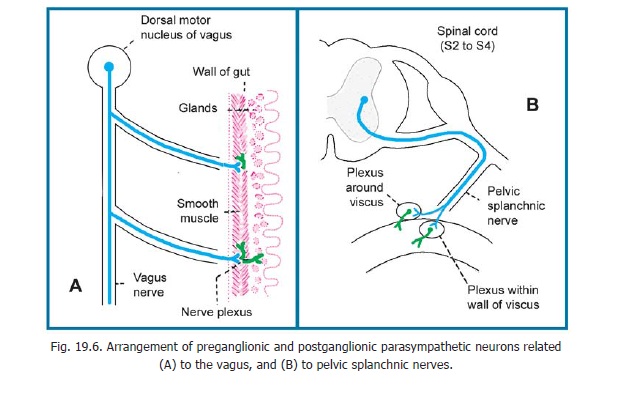
1. The first group is located in the general visceral efferent nuclei of the brainstem. Axons arising in these nuclei constitute the cranial parasympathetic outflow (Fig. 19.3). They pass through the third, seventh, ninth and tenth cranial nerves to terminate in peripheral ganglia. The largest part of this outflow is constituted by the vagus nerve. Its fibres terminate in relation to postganglionic neurons located in thoracic and abdominal autonomic plexuses.
2. The second group of parasympathetic preganglionic neurons is located in the second, third and fourth sacral segments of the spinal cord (Fig. 19.3). Their axons constitute thesacralparasympathetic outflow. They emerge from the cord through the anterior nerve roots of thecorresponding spinal nerves. The axons leave the spinal nerves to form the pelvic splanchnicnerves which end in pelvic autonomic plexuses (Fig. 19.6B).
Parasympathetic Postganglionic Neurons
1. Postganglionic neurons related to the third, seventh and ninth cranial nerves are located in the ciliary, submandibular, pterygopalatine and otic ganglia. Some subsidiary ganglia may be located in the vicinity of these ganglia.
2. Postganglionic neurons related to the vagus are located in thoracic and abdominal autonomic plexuses, close to, or within, the viscera supplied (Fig. 19.6). The axons arising from these postganglionic neurons innervate various thoracic and abdominal viscera including the greater part of the gut.
3. Postganglionic neurons related to the sacral parasympathetic outflow are located in pelvic autonomic plexuses. They innervate the pelvic viscera. They also supply the rectum, the sigmoid colon, the descending colon, and the left one third of the transverse colon. Like the postganglionic neurons related to the vagus, these neurons are located close to, or within, the viscera supplied, their axons running a short course to supply the viscus concerned.
Related Topics