Chapter: Human Nervous System and Sensory Organs : Spinal Cord and Spinal Nerves
Posterior Fascicle - Infraclavicular Part of Peripheral Nerves
Posterior Fascicle
The posterior fascicle gives rise to the axil-lary nerve and the radial nerve.
Axillary nerve (C5 – C6).This runs deep in-side the axilla and across the capsule of the shoulder joint around the surgical neck on the back of the humerus. It passes through the lateral axillary gap and extends beneath the deltoid muscle to the anterior margin of the latter.
Before the nerve trunk passes through the lateral axillary gap, it gives off a motor branch (DF3) to the lesser teres muscle (D4), which also passes through the lateral axil-lary gap. At the same level, the superiorlateral cutaneous nerve of the arm (D–F5)branches off and reaches the skin at the pos-terior margin of the deltoid muscle, where it supplies the skin of the lateral aspects of shoulder and upper arm. From the nerve trunk extending beneath the deltoid muscle to the front, numerous branches (D6) to the deltoid muscle (D7) branch off and supply its various parts.
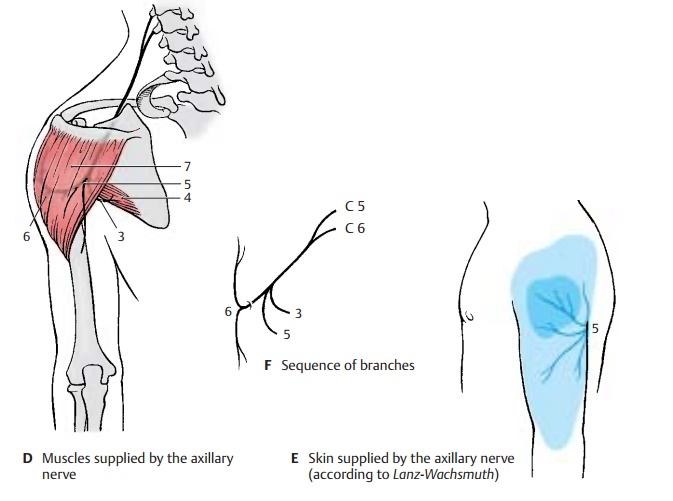
Clinical Note: As a result of its location on thecapsule of the shoulder joint, the nerve can be in-jured by dislocation of the humerus or by humeral neck fracture. This causes anesthesia in the skin area over the deltoid muscle.
Innervation of the skin (B, C, E).Auto-nomic zone (dark blue) and maximum zone (light blue).
Radial nerve (C5 – C8) (A – C). The main nerve of the posterior cord supplies the ex- tensor muscles of upper arm and forearm.
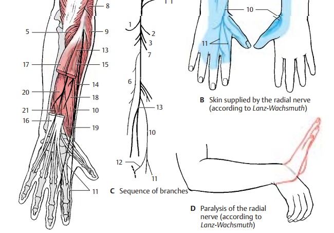
The nerve trunk extends from the axilla into the proximal third of the medial bicipital sulcus and then spirals around the dorsal surface of the humerus, to which it is directly apposed in the sulcus of the radial nerve. In the distal third of the upper arm, it passes to the flexor side between brachial muscle and brachioradial muscle. In the sul- cus of the radial nerve, the nerve can easily be injured by pressure or by bone fractures because of its proximity to the bone. The nerve crosses the elbow joint on the flexor side and divides at the level of the head of radius into two terminal branches, the su-perficial branch and the deep branch. The su- perficial branch continues in the forearm on the medial surface of the brachioradial muscle and then runs in the lower third be- tween brachioradial muscle and radius to the extensor side in order to reach the back of the hand. The deep branch obliquely penetrates the supinator muscle, gives off numerous muscular branches, and extends as the thin posterior interosseous nerve of the forearm to the wrist.
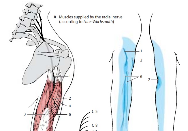
For the upper arm, the radial nerve gives off the posterior cutaneous nerve of the arm (A – C1), which supplies a skin area on the extensor side of the upper arm with sensory fibers, and the inferior lateral cutaneous nerve of the arm (A – C2). In the middle third of the upper arm, it gives off muscular branches (AC3) for the long head, the lateral head, andthe medial head of the triceps muscle (A4). The branch for the medial head gives off also the branch for the anconeus muscle (A5).
The posterior cutaneous nerve of the forearm(A – C6) branches off in the region of thethen ramifies into its two major branches in the forearm. At the back of the hand, the superficial branch (A – C10) gives off the dorsal digital nerves (A – C11); they supply sensory fibers to the radial back of the hand, the extensor side of the thumb, the proximal phalanges of index and middle fingers, and the radial half of the extensor side of the ring finger. The ulnar communicating branch of the radial nerve connects with the ulnar nerve (C12).
The deep branch (AC13) gives off muscular branches to the short radial extensor muscle of the wrist (A14) and to the supinator muscle, while passing through the supina- tor muscle. Thereafter, it gives off motor branches to the hand extensor muscles, namely, to the common extensor muscle of the fingers (A15), the extensor muscle of the little finger (A16), the ulnar extensor muscleof the wrist (A17), the long abductor muscle of the thumb (A18), and the short extensor muscle of the thumb (A19). Finally, the ter- minal branch of the deep branch, the poste- rior interosseous nerve, gives off branches to the long extensor muscle of the thumb (A20) and to the extensor muscle of the index finger (A21).
The nerve sends sensory branches to the shoulder joint and wrist.
Clinical Note: Injury to the main nerve trunk in the area of the upper arm results in paralysis of the extensor muscles. This mainly affects the hand, leading to the so-called wristdrop (D) characteristic for radial paralysis: extension is possible neither in the wrist nor in the fingers, thus making the hand drop down limply.
Innervation of the skin (B).Autonomic zone (dark blue) and maximum zone (light blue).
Related Topics