Sexual Reproduction in Plants - Post Fertilization structure and events | 12th Botany : Chapter 1 : Asexual and Sexual Reproduction in Plants
Chapter: 12th Botany : Chapter 1 : Asexual and Sexual Reproduction in Plants
Post Fertilization structure and events
Post
Fertilization structure and events
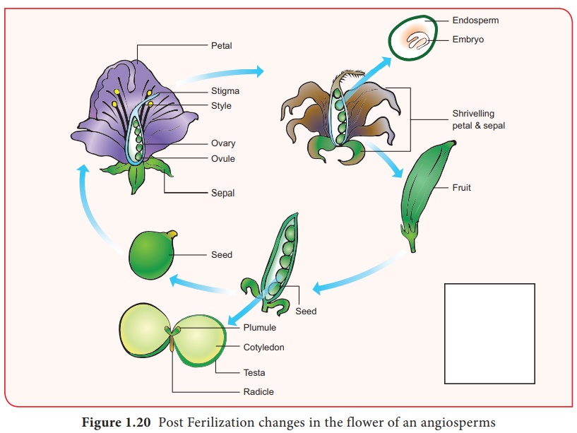
After fertilization,
several changes take place in the floral parts up to the formation of the seed
(Figure 1.20).
The events after
fertilization (endosperm, embryo development, formation of seed, fruits) are
called post fertilization changes.
Endosperm
The primary endosperm
nucleus (PEN) divides immediately after fertilization but before the zygote
starts to divide, into an endosperm. The primary endosperm nucleus is the
result of triple fusion (two polar nuclei and one sperm nucleus) and thus has
3n number of chromosomes. It is a nutritive tissue and regulatory structure
that nourishes the developing embryo. Depending upon the mode of development
three types of endosperm are recognized in angiosperms. They are nuclear
endosperm, cellular endosperm and helobial endosperm (Figure 1.21).
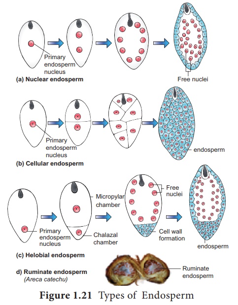
Nuclear endosperm: In the nuclear type, the Primary endosperm nucleus (PEN) divides into two without any wall formation. The subsequent division of these two nuclei are free nuclear so that the endosperm consists of only free nuclei and cytoplasm around them. The nuclei may either remain free or may become separate by walls in later stages. Examples: Coccinia, Capsella and Arachis.
Cellular endosperm: In this type the primary
endosperm nucleus(PEN) divides into 2 nuclei which are immediately followed
by a wall formation. Subsequent divisions are also followed by walls. Examples:
Adoxa, Helianthus and Scoparia.
Helobial endosperm: In helobial type, the
primary endosperm nucleus (PEN) moves towards the base of the embryo sac
where it divides into two nuclei. These 2 nuclei are separated by a wall to
form a large micropylar chamber and a small chalazal chamber. The nucleus of
the micropylar chamber undergoes several free nuclear divisions whereas that of
the chalazal chamber may or may not divide. Examples : Hydrilla and
Vallisneria
The endosperms may
either be completely consumed by the developing embryo or it may persist in the
mature seeds. Those seeds without endosperms are called non- endospermous or
ex- albuminous seeds. Examples: Pea, Groundnut and Beans. Those seeds
with endosperms are called endospermous or albuminous seeds. The endosperms in
these seeds supply nutrition to the embryo during seed germination. Examples:
Paddy, Coconut and Castor.
Ruminate endosperm: The endosperm with irregularity
and unevenness in its surface forms ruminate endosperm (Example: Areca
catechu). The activity of the seed coat or the endosperm itself results in
this type of endosperm. The unequal radial elongation of the layer of seed coat
results in the rumination of endosperm in Passiflora. In Annonaceae and
Aristolochiaceae definite ingrowth or infolding of the seed coat produces
ruminate endosperm. The irregular surface of the seed coat makes endosperm
ruminate in Myristica.
Functions of endosperm:
·
It is the nutritive tissue for the developing embryo.
·
In majority of angiosperms, the zygote divides only after the
development of endosperm.
·
Endosperm regulates the precise mode of embryo development.
Endosperm haustoria
Another interesting
feature of the endosperm is the presence of haustoria. In the case of helobial
endosperm the chalazal chamber itself acts as a haustorial structure.
In cellular and nuclear
endosperm special structures are produced towards the micropylar, chalazal,
both micropylar and chalazal which may be in lateral direction depending on the
species. These absorb nutrients from other outer tissue or from ovary tissue
and supply them to the growing embryo.
Embryogenesis
Development of Dicot embryo
The development of Dicot
embryo (Capsella bursa-pastoris) is of Onagrad or crucifer type.
The embryo develops at micropylar end of embryo sac.
The Zygote divides by a
transverse division forming upper or terminal cell and lower or basal
cell. The basal cell divides transversely and the terminal cell
divides vertically to form a four celled proembryo. A second vertical division
right angle to the first one takes place in terminal cell forming a four celled
stage called quadrant. A transverse division in the quadrant results in
eight cells arranged in two tiers of four each called octant stage.
The upper tier of four
cells of the octant is called epibasal or anterior octant and the lower
tier of four cells constitute hypobasal or posterior octants. A
periclinal division in the octants results in the formation of 16 celled stage
with eight cells in the outer and eight in the inner.
The outer eight cells
represent the dermatogen and undergoes anticlinal division to produce
epidermis. The inner eight cells divide by vertical and transverse division to
form outer layer of periblem which give rise to cortex and a central
region of pleurome which forms stele
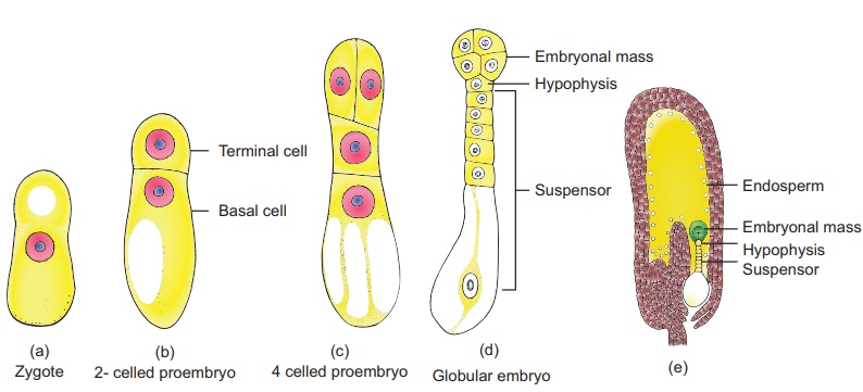
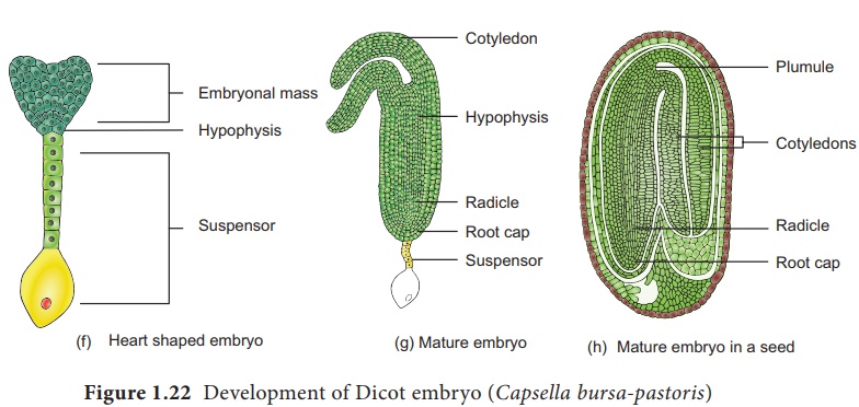
During the development, the two cells of the basal cell undergoes several transverse division to form a six to ten celled suspensor. The embryo at this stage become globular and the suspensor helps to push the embryo deep into the endosperm. The uppermost cell of the suspensor enlarge to form a haustorium. The lowermost cell of the suspensor is called hypophysis. A transverse division and two vertical division right angle to each other of hypophysis results in the formation of eight cells. The eight cells are arranged in two tiers of four cells each The upper tier give rise to root cap and epidermis. At this stage embryo proper appears heart shaped, cell divisions in the hypocotyl and cotyledon regions of the embryo proper results in elongation. Further development results in curved horse shoe shaped embryo in the embryo sac. The mature embryo has a radicle, two cotyledons and a plumule(Figure 1.22).
Seed
The fertilized ovule is
called seed and possesses an embryo, endosperm and a protective coat. Seeds may
be endospermous (wheat, maize, barley and sunflower) or non endospermous.
(Bean, Mango, Orchids and cucurbits).
Structure of a Cicer seed as an example for Dicot seed
The mature seeds are
attached to the fruit wall by a stalk called funiculus. The funiculus
disappears leaving a scar called hilum. Below the hilum a small pore
called micropyle is present. It facilitates entry of oxygen and water
into the seeds during germination. Each seed has a thick outer covering called
seed coat. The seed coat is developed from integuments of the ovule. The outer
coat is called testa and is hard whereas the inner coat is thin,
membranous and is called tegmen. In Pea plant the tegmen and testa are
fused. Two cotyledons laterally attached to the embryonic axis are present. It
stores the food materials in pea whereas in other seeds like castor the
endosperm contains reserve food and the cotyledons are thin. The portion of
embryonal
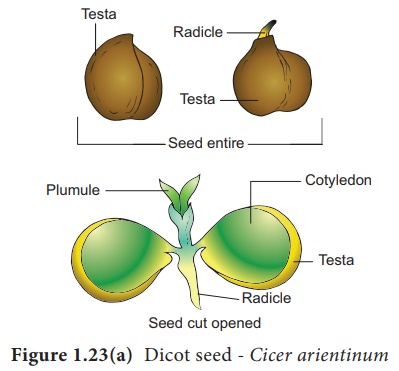
The other end of the
axis called embryonic shoot is the plumule. Embryonal axis above the
level of cotyledon is called epicotyl whereas the cylindrical region
between the level of cotyledon is called hypocotyl(Figure 1.23 a). The
epicotyl terminates in plumule whereas the hypocotyl ends in radicle.
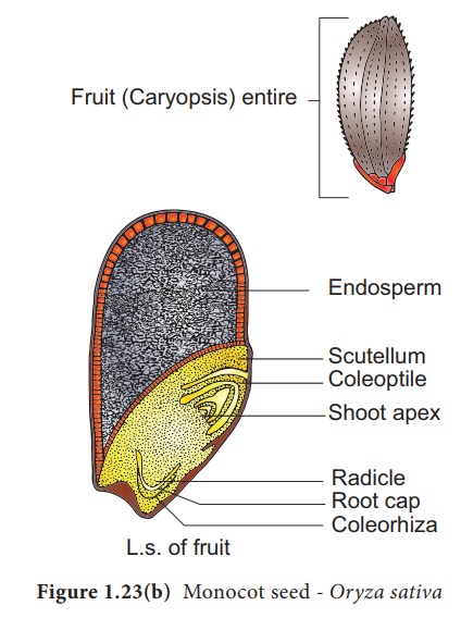
Structure of Oryza seed as an example for Monocot seed
The seed of paddy is one
seeded and is called Caryopsis. Each seed remains enclosed by a
brownish husk which consists of glumes arranged in two rows. The seed coat is a
brownish, membranous layer closely adhered to the grain. Endosperm forms the
bulk of the grain and is the storage tissue. It is separated from embryo by a
definite layer called epithelium. The embryo is small and consists of
one shield-shaped cotyledon known as scutellum present towards lateral
side of embryonal axis. A short axis with plumule and radicle protected by the root
cap is present. The plumule is surrounded by a protective sheath
called coleoptile. The radicle including root cap is also covered by a
protective sheath called coleorhiza. The scutellum supplies the growing
embryo with food material absorbed from the endosperm with the help of the
epithelium (Figure 1.23 b).
Related Topics