Classification, External features, Exoskeleton, Endoskeleton, Anatomy Structure - Pigeon (Columba livia) | 11th Zoology : Chapter 4 : Organ and Organ Systems in Animals
Chapter: 11th Zoology : Chapter 4 : Organ and Organ Systems in Animals
Pigeon (Columba livia)
Pigeon - Columba livia
Birds
belong to the Class Aves (L. avis -
birds). The most distinguishing feature of birds is the possession of feathers.
The study of birds is Ornithology. A
bird is a feathered, bipedal, flying
vertebrate possessing wings. Their
external and internal organization
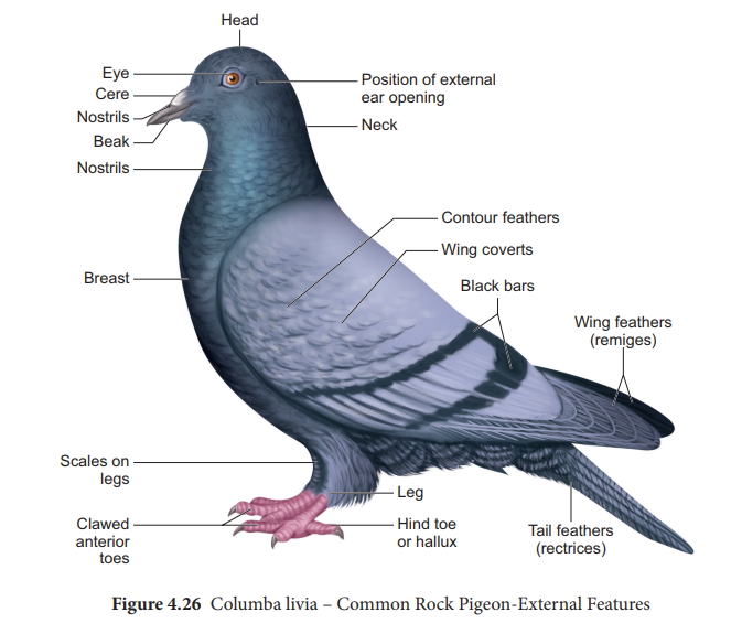
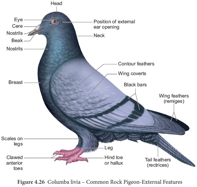
correlates with its aerial habit. More than 500 species of pigeon exist
throughout the world. In India, about 10 species of pigeons are found. Columba
livia is found throughout India (Figure 4.26).
Classification
Phylum : Chordata
Class : Aves
Order : Columbiformes
Genus : Columba
Species : livia
External features
The
compact, boat shaped streamlined body of pigeon is well adapted for their
aerial mode of life. The body of pigeon is
Head
is comparatively small, spherical and situated at the anterior most part of the
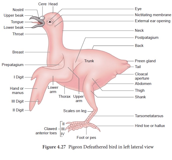
body. Beaks present anteriorly are
formed by the elongation of upper and lower jaw and they are devoid of teeth.
At the base of the beak are the external
nostrils overhung by a swollen, sensitive soft skin called cere. Eyes are prominent, round and laterally present. Eyes are protected
by an upper eyelid, lower eyelid,
and a transparent nictitating membrane.
Posterior to the eyes are the ear
openings which lead to the tympanic
membrane by short tube, external
auditory meatus. Neck is flexible, cylindrical and long which connects the head
with the trunk. The spindle shaped trunk bears a pair of wings and a pair of legs. The cloacal aperture opens ventrally at the hind end of the trunk.
Dorsally the base of the tail has a
knob like papilla, which bears the opening of the preen gland or uropygial
gland. It is the only cutaneous gland present and its oily secretion is
used for lubricating or preening the feathers. The tail is used as a rudder in
flight. Fore limbs are modified into
wings. The wings have three typical regions, the upper arm (brachium), lower
arm (ante - brachium) and the hand (manus). Three clawless and imperfectly
marked digits are present on each hand. While at rest, each forelimb is folded
in the form of ‘Z’; during flight
they are extended. With the modification of the forelimbs for flight, the whole
weight of the body is supported by the hind
limbs , while the bird is at rest or walking; the hind limbs are therefore
attached anteriorly from the trunk to balance the body and support the weight
of the body at rest. They are warm blooded or homeothermic.
Exoskeleton
The
exoskeleton of pigeon is derived from the epidermis
and occurs in the form of horny claws,
scales and feathers. Beaks are
used for ingestion, fighting and preening
of feathers. Claws are used for walking and perching. Epidermal scales are
present on the foot and the entire body is covered by feathers. Arrangement of
feathers on the body of bird is called pterylosis
. Feathers are of three kinds: large quill
feathers on wings and tail which are used for flight; contour feathers, form a covering for the body and filoplumes, lie between the contour
feathers. The nestlings are covered
with down feathers which resemble
the filoplumes.
Structure of a Quill feather
The quill
feather has a stem or scapus and is divided into a lower
hollow part called calamus or quill and an upper solid portion
called rachis. Lower end of the stem
has an opening called inferior
umbilicus which receives a dermal papilla, supplying nutrients and pigments
for the growing feathers (Figure 4.28)
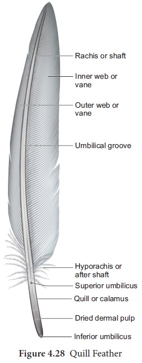
A second opening the superior umbi-licus occurs at the junction of the quill and the rachis, on the inner face of the feather; close to this opening is a small tuft of soft feathers called after shaft . Attached to the rachis are small filament or barbs ; the ra-chis with the barbs constitute the vane or the vexillum. Each barb is fringed with an oblique set of processes called barbules, which have minute hooklets or barbi-cels by which adjacent barbs are hooked together to form a continuous blade for striking the air during flight.
Anatomy
Endoskeleton
The
skeletal system is strong but lightly built. The bones are light and spongy.
Many of the long bones contain air instead of marrow (Pneumatic bones). This
reduces the weight of the body. The breast
bone or sternum has a broad
plate of bone produced ventrally into a prominent vertical crest or keel to which
the powerful muscles of flight are attached.
Flight muscles
Wings are modified forelimbs and the organs of flight. The musculature of the forelimbs are greatly modified in response to the function they perform. Flight is the coordinated effort of a number of paired muscles. The muscles which operate the wings during flight are called flight muscles. The major flight muscles of pigeon are the pectoral muscles. Pectoral muscles are of two types namely the Pectoralis major and Pectoralis minor. The pectoralis major muscle is a large and powerful flight muscle which arises from the sternum.
Contraction of these muscles lower
the wings in flight. Pectoralis minor (subclavius) is small and elongated
muscle which elevates the wings during flight. Besides the pectoralis, the
small coracobrachialis muscle also
helps to pull the wings down and to rotate wings during flight.
Digestive System
The long
coiled alimentary canal consist of buccal cavity, pharynx, oesophagus, crop,
stomach, small intestine and Large intestine. (Figure 4.29).
Mouth is
covered by a toothless, horny, upper and lower beaks. Behind the mouth, there is a wide buccal cavity.
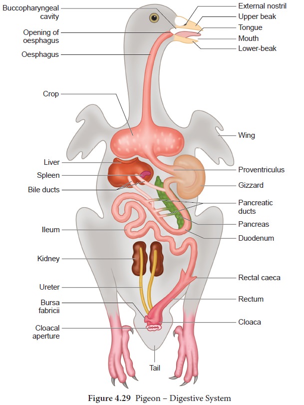
In the
floor of the buccal cavity, a large,
narrow, horny tongue is present with
scanty sensory papillae and numerous mucus glands. Buccal cavity leads into
the pharynx fol-lowed by the oesophagus, which enlarges to form a thin walled,
bilobed elastic sac, the crop. The
crop serves as a food res-ervoir. Beyond the crop the oesophagus enters the
stomach which is differentiat-ed into anterior glandular proventriculus and a posterior muscular ventriculus or gizzard.
The proventriculus has a mucus lining which secretes the gastric juice. The
walls of the gizzard is thick, muscular and has many tubular glands. The cavity
of the gizzard contains grit or small peb-bles called gastroliths that are swallowed by the bird. These stones helps the
bird in grinding the food. The gizzard leads to a small intestine which consists of a ‘U’ shaped duodenum and ileum. The pan-creas lies between the two limbs of
the duodenum and receives three ducts from the pancreas and two bile ducts from
the liver. The inner lining of the
ileum con-tains numerous villi which
helps in ab-sorption. The ileum continues into the large intestine, which is short and is dif-ferentiated into
rectum and cloaca. A pair of small blind pouches called rectal caeca is present at the junction of the ileum and rectum.
The rectum leads into the cloaca
which is divided into the anterior copro-daeum
into which the rectum opens, the middle urodaeum
into which the urini-genital ducts
open, and the posterior ves-tibule
or proctodaeum, which opens to the
outside by the cloacal aperture.
Buccal
glands, salivary glands, gas-tric glands, liver, pancreas and intestinal glands
are the digestive glands which
en-hance the process of digestion in pigeon. There is no gall bladder in the pigeon though present in many other birds.
Pi-geons produce ‘milk’, a cheesy
and nour-ishing secretion, from both the sexes. It is formed by the
degeneration of the epithe-lial cells lining the crop. It is regurgitated and
fed to the young birds.
The pigeon feeds on grains. As birds have no teeth, the food swallowed by it passes through the gullet or oesophagus into the crop where it is stored. There are mucous glands in the crop; food is softened by being mixed with the mucus and the se-cretion of the buccal glands, aided by the warmth of the body. The food then enters the stomach, where it is digested by gastric juices secreted in the proventriculus; the food is also crushed in the gizzard with aid of gastroliths. The food is thus reduced to smaller particles and the partly digested food passes into the intestine where it is mixed with the bile and pancreatic juice, and further digestion is effected.
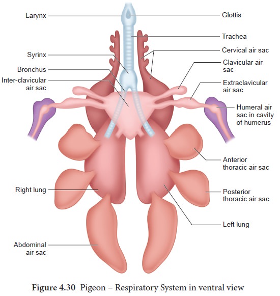
Respiratory system
In birds
the type of respiration is pulmonary.
The respiratory system includes the respiratory tract, the respiratory organs
and air sacs. A true muscular diaphragm is absent in birds.
The respiratory tract includes the
nares, nasal sacs, glottis, larynx, trachea and syrinx. The respiratory organs are the lungs and
air sacs. The larynx opens into the trachea and is supported by a series of
closely set rings. The trachea divides into two bronchi, each of which divides and sub-divides into smaller
branches, ultimately ending in fine air-capillaries which lies intermingled
with the capillaries of the pulmonary vessels. Lungs are solid spongy organs;
attached dorsally to the ribs. There are nine air-sacs: a pair of cervical sacs
at the base of the neck one on each side; a single median interclavicular air
sac connected with both lungs and situated in between the two limbs of the
furcula and on either sides it gives off an extraclavicular air sac
communicating with an air - cavity of the humerus and a clavicular air sac; two
pairs of thoracic air sacs and a pair of abdominal air sacs. This complicated
arrangement adds to the efficient respiratory function and maintenance of a
high temperature (Figure 4.30).
Respiratory mechanism
The lungs
are not dilatable since the skeleton around them forms a rigid framework.
Inspiration is passive and expiration is an active process. During respiration
the sternum is drawn towards the vertebral column, by contraction of the
muscles of the body-wall. As is drawn up, the elastic ribs are bent so as to
bring about a decrease in the size of the body cavity and the air from the
lungs is forced out. When the muscles relax, the body-cavity recovers its size
and air is drawn in.
Syrinx
The
larynx does not take part in the production of voice. The voice box lies deep down where the trachea divides into two
bronchi, and is known as syrinx, a
structure characteristic of birds. It consists of a chamber with its walls
supported by three or four rings of the trachea and the first ring of each
bronchus; its inner lining is raised into folds, the vibrations of which is
caused by the movement of air results in the production of sound.
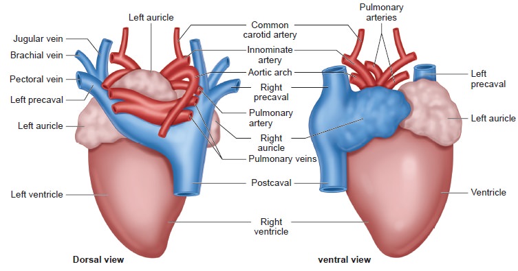
Circulatory system
Pigeon
has an efficient circulatory system to meet the metabolic demands of flight,
but also plays a significant role in maintaining the body temperature. The
circulatory system of pigeon includes the heart and blood vessels. The heart of the pigeon is four chambered with two auricles and two ventricles. There is no sinus
venosus. The two precaval veins or superior venae cavae, a post caval vein or inferior vena cava opens into the right auricle; the pulmonary
aorta and systemic trunks arise from the right and left ventricles
respectively. The right side of the heart is completely separated from the left
side of the heart by a septum. The right auricle opens into the right ventricle
by the right auriculo -ventricular
aperture and the left auricle into the left ventricle by the
left auriculo- ventricular aperture.
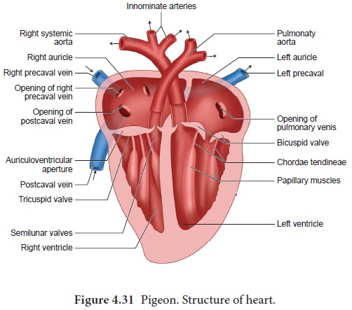
There are valves at these apertures,
which allows the blood to flow only in one direction, i.e., from the auricle
into the ventricle but not backwards. The right auriculo-ventricular valve
consists of a single flap without connecting chordae tendinae; the valve on the left side has two flaps
connected to the papillary muscles
by chordae tendinae. The pulmonary aorta arises from the right ventricle and
the aortic arch from the left ventricle. The pulmonary veins open into the left
auricle. There are three semilunar
valves at the junction of the pulmonary aorta and the right ventricle. The
pulmonary aorta divides into two branches, each entering a lung. Only the right
aortic arch is present in birds.
The right
auricles of the heart receives venous blood from all parts of the body except
the lungs, through the precaval and post caval veins. The right ventri-cles
pumps venous blood into the lungs through the pulmonary aorta. The oxy-genated blood from the lungs is returned
to the left auricle through the pulmonary
veins. From the left ventricle a single right aortic arch carries
oxygenated blood to the different parts of the body. The right half of the
heart receives and discharges only venous
blood and the left half only arterial
blood. Thus birds possess a com-plete
double circulation which includes the pulmonary
circulation and systemic circulation.
(Figure 4.31).
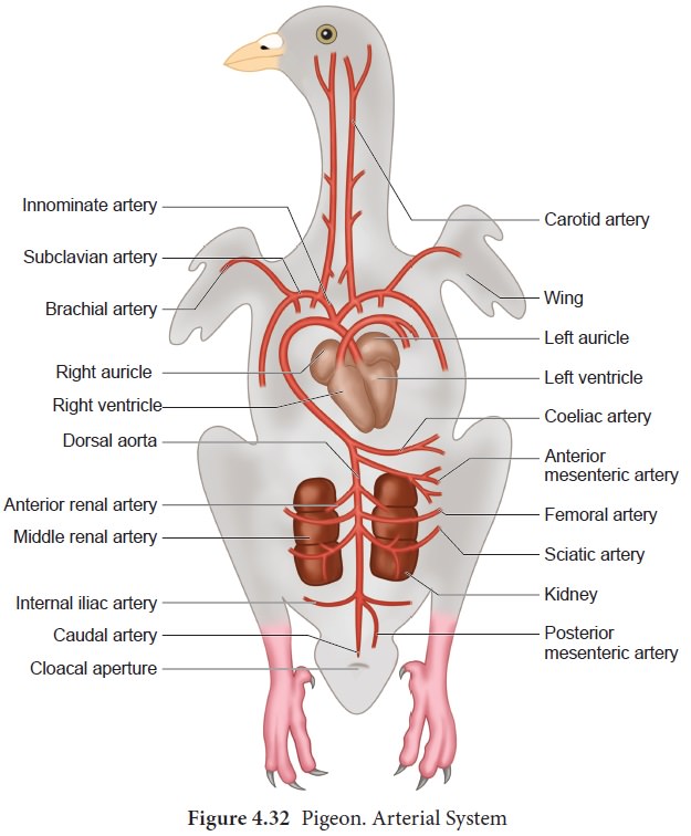
The arterial system
The right aortic arch curves over to the right side giving off at the curve the right and the left innominate arteries; each of these gives rise to a carotid artery and a subclavian artery, the former carrying blood to the brain.
The subclavian artery divides into a brachial artery conveying blood to the arm, and a pectoral artery to the muscles of the wings. The aortic arch passes backwards as the dorsal aorta, from which are given off the unpaired coeliac artery supplying blood to the stomach, the liver and few parts of intestine; the unpaired anterior mesenteric artery to the great part of the intestine; the paired anterior renal arteries to the anterior lobes of the kidney; the paired femoral arteries supplying blood to the anterior region of the thigh and the paired sciatic arteries supply blood to the posterior parts of the thighs and the leg.
From the each sciatic artery arises a middle renal artery to the middle lobe of the kidney
and a posterior renal artery to the posterior lobe; the unpaired posterior
mesenteric artery supplies blood to
the rectum and the cloaca; the paired internal
iliac arteries to the pelvis and the caudal
artery which is the terminal portion of the dorsal aorta extends to the
tail (Figure 4.31).
The venous system
The precaval vein of each side is formed by the union of the jugular vein from the head, the brachial vein from the arm, and the pectoral vein from the pectoral muscles. The jugular vein of the two sides are connected in front by a transverse vessel. The postcaval vein is formed by the union of the two iliac veins in front of the kidney. Each iliac vein is in turn formed by the union of the femoral vein from the leg, an efferent renal vein from the kidney, and the renal-portal vein from the posterior regions. The hepatic-portal circulation is present and the blood from the liver is emptied into the postcaval vein by three hepatic veins. (Figure 4.33).
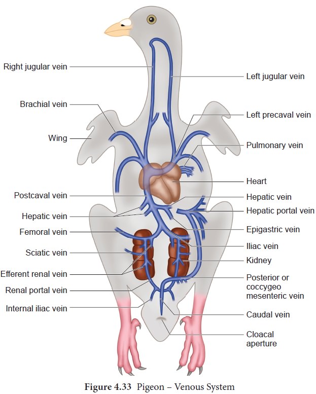
The caudal vein from the tail divides into
the right and the left renal-portal vein each of which enters the kidney.
Be-fore entry, the renal-portal vein is joined by the internal iliac vein from the pelvis. As the renal-portal vein passes through the kidney, it receives the sciatic and the femoral vein from the leg,
and final ly emerges from the kidneys as the iliac vein. The renal- portal
veins do not break into capillaries in the kidney but only send a few small
branches; renal -portal circulation is therefore not well devel-oped in the
bird.
At the place of bifurcation of the cau-dal vein into the two renal-portal veins arises the median coccygeomesenter-ic vein which is characteristic of birds.
This vein
runs forward, receives in its course veins from the rectum, and joins the hepatic portal vein. The epigastric vein returns the blood from
the mesen-teries and joins one of
the hepatic veins.
Nervous system and receptor organs
The
nervous system consists of central
nervous system which includes the brain and spinal cord, the peripheral nervous system and the autonomous nervous system (Figure
4.34). The brain of pigeon is larger than in lower forms, it is short, broad
and rounded within cranial cavity. It is covered by two meninges, the outer duramater
and an inner pia- arachnoid membrane
and the space between the two meninges is filled with cerebrospinal fluid. The cerebral
hemispheres of the pigeon are large and extend behind to meet the cerebellum. The cerebrum controls
voluntary movements and is the centre for memory and intelligence. The diencephalon is covered dorsally by the
cerebral hemispheres and cerebellum. The diencephalon relays impulses to the
cerebral hemispheres, integrates the autonomic
system and the perception of extreme cold, pain, heat etc. On the ventral side
of the diencephalon is the optic
chiasma, behind the chiasma projects the infundibulum bearing a large hypophysis
or pituitary. The
optic lobes are large and occupy a lateral position owing to the large size of
the cerebral hemispheres and cerebellum. Optic lobes are centres for sight. The
pineal body and infundibulum are present. The cerebellum is highly developed
and convoluted indicating the delicate sense of equilibrium and the great power
of muscular co-ordination required for birds.
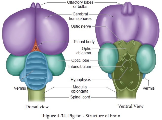
The
cerebellum extends backwards covering a large part of the medulla oblongata
which descends downwards to join the spinal
cord. The medulla oblongata controls the involuntary movement. The
olfactory lobes or bulbs are small and degenerate due to poorly developed
organs of smell.
The
peripheral nervous system consists of 12 pairs of cranial nerves and 38 pairs of spinal
nerves. The autonomic nervous system of pigeon includes the sympathet-ic and parasympathetic nervous system. It contains the nerves and
ganglia. The sympa-thetic nerves
supply the alimentary, respira-tory, circulatory and urinogenital systems.
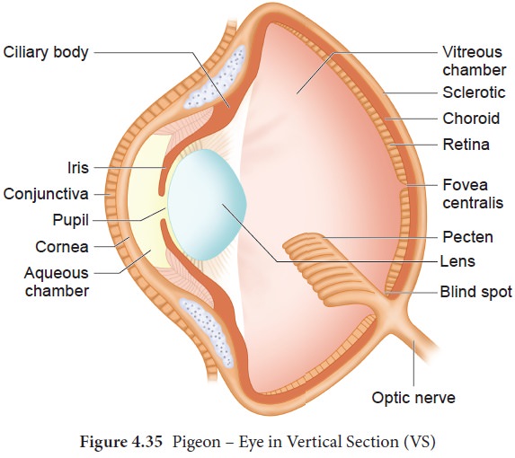
Sense organs
Eyes are large and well developed;
they are not spherical, but biconvex. The sclerotic coat contains bony plates. There is a vascular pigmented plaited
process known as the pecten, projecting
into the vitreous body from the
point where the optic nerve enters the
eye (Figure 4.35). Pecten is concerned with the power of accommodation which is
greatly developed in birds. The muscles for the movement of the eye-balls are
reduced. In the ear, the cochlea is well developed. The two eustachian tubes unite and open by a
common aperture on the roof of the buccal cavity. The olfactory sense is poorly developed.
Urinogenital system
Excretory System
The
paired kidneys which are metanephric
are flat, elongated and lobulated. The ureters lead directly backward to open
into the urodaeum or middle
compartment of the cloaca; there is no urinary bladder. The nitrogenenous waste
is excreted in the form of uric acid and discharged as a semi-solid mass. Adrenal bodies lie attached to the
ventral surface of the kidneys as small yellowish elongated streaks.
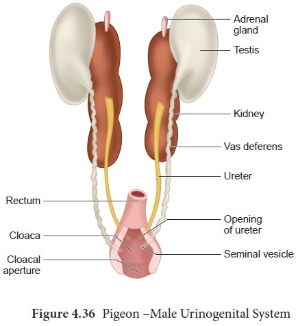
Reproductive system.
A pair of
ovoid testes are attached to anterior
end of the kidneys by peritoneum (Figure 4.36). From each testis leads the vas deferens which runs backwards along
the outer side of the ureter of that side, and opens on a small papilla into
the urodaeum. The vas deferens is dilated into a seminal vesicle at its hind end. There is no copulatory organ.
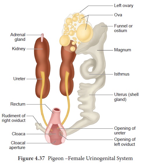
The female reproductive organs consist of a
single ovary on the left side which
is an adaptation to aerial life and
an oviduct which opens into the body-cavity by a funnel-like aperture at the
anterior end and posteriorly opens into the urodaeum (Figure 4.37).
Related Topics