Chapter: Medical Physiology: The Liver as an Organ
Physiologic Anatomy of the Liver
Physiologic Anatomy of the Liver
The liver is the largest organ in the body, contributing about 2 per cent of the total body weight, or about 1.5 kg in the average adult human. The basic functional unit of the liver is the liver lobule, which is a cylindrical structure several millimeters in length and 0.8 to 2 millimeters in diameter. The human liver contains 50,000 to 100,000 individual lobules.
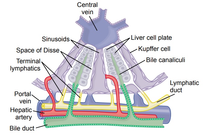
The liver lobule, shown in cut-away format in Figure 70–1, is constructed around a central vein that empties into the hepatic veins and then into the vena cava. The lobule itself is composed principally of many liver cellular plates (two of which are shown in Figure 70–1) that radiate from the central vein like spokes in a wheel. Each hepatic plate is usually two cells thick, and between the adjacent cells lie small bilecanaliculi that empty into bile ducts in the fibrous septa separating the adjacent liverlobules.
In the septa are small portal venules that receive their blood mainly from the venous outflow of the gastrointestinal tract by way of the portal vein. From these venules blood flows into flat, branching hepatic sinusoids that lie between the hepatic plates and then into the central vein. Thus, the hepatic cells are exposed con-tinuously to portal venous blood.
Hepatic arterioles are also present in the interlobular septa. These arteriolessupply arterial blood to the septal tissues between the adjacent lobules, and many of the small arterioles also empty directly into the hepatic sinusoids, most frequently emptying into those located about one third the distance from the interlobular septa, as shown in Figure 70–1.
In addition to the hepatic cells, the venous sinusoids are lined by two other types of cell: (1) typical endothelial cells and (2) large Kupffer cells (also called reticu-loendothelial cells), which are resident macrophages that line the sinusoids and are capable of phagocytizing bacteria and other foreign matter in the hepatic sinus blood.
The endothelial lining of the sinusoids has extremely large pores, some of which are almost 1 micrometer in diameter. Beneath this lining, lying between the endothelial cells and the hepatic cells, are narrow tissue spaces called the spaces ofDisse, also known as the perisinusoidal spaces. The millions of spaces of Disseconnect with lymphatic vessels in the interlobular septa. Therefore, excess fluid in these spaces is removed through the lymphatics. Because of the large pores in the endothelium, substances in the plasma move freely into the spaces of Disse. Even large portions of the plasma proteins diffuse freely into these spaces.
Related Topics