Chapter: Medical Physiology: Cerebral Cortex, Intellectual Functions of the Brain, Learning and Memory
Physiologic Anatomy of the Cerebral Cortex
Physiologic Anatomy of the Cerebral Cortex
The functional part of the cerebral cortex is a thin layer of neurons covering the surface of all the convolutions of the cerebrum. This layer is only 2 to 5 millimeters thick, with a total area of about one quarter of a square meter. The total cerebral cortex contains about 100 billion neurons.
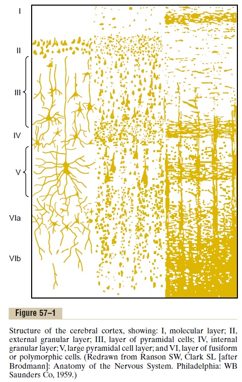
Figure 57–1 shows the typical histological structure of the neuronal surface of the cerebral cortex, with its successive layers of different types of neurons. Most of the neurons are of three types: (1) granular (also called stellate), (2) fusiform, and (3) pyramidal, the last named for their characteristic pyramidal shape.
The granular neurons generally have short axons and, therefore, function mainly as interneurons that transmit neural signals only short distances within the cortex itself. Some are excitatory, releasing mainly the excitatory neurotransmitter gluta-mate; others are inhibitory and release mainly the inhibitory neurotransmitter gamma-aminobutyric acid (GABA). The sensory areas of the cortex as well as theassociation areas between sensory and motor areas have large concentrations of these granule cells, suggesting a high degree of intracortical processing of incoming sensory signals within the sensory areas and association areas.
The pyramidal and fusiform cells give rise to almost all the output fibers from the cortex. The pyramidal cells are larger and more numerous than the fusiform cells. They are the source of the long, large nerve fibers that go all the way to the spinal cord. They also give rise to most of the large subcortical association fiber bundles that pass from one major part of the brain to another.
To the right in Figure 57–1 is shown the typical organization of nerve fibers within the different layers of the cerebral cortex. Note particularly the large number of horizontal fibers that extend between adjacent areas of the cortex, but note also the vertical fibers that extend to and from the cortex to lower areas of the brain andsome all the way to the spinal cord or to distant regions of the cerebral cortex through long association bundles.
The functions of the specific layers of the cerebral cortex are discussed in Chap-ters 47 and 51. By way of review, let us recall that most incoming specific sensory signals from the body terminate in cortical layer IV. Most of the output signals leave the cortex through neurons located in layers V and VI; the very large fibers to the brain stem and cord arise generally in layer V; and the tremendous numbers of fibers to the thalamus arise in layer VI. Layers I, II, and III perform most of the intra-cortical association functions, with especially large numbers of neurons in layers II and III making short horizontal connections with adjacent cortical areas.
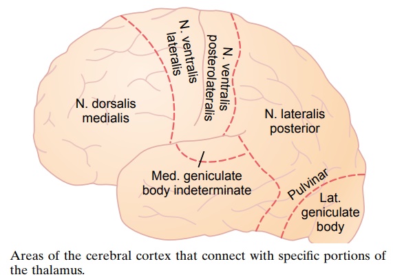
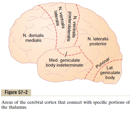
Anatomical and Functional Relations of the Cerebral Cortex to the Thalamus and Other Lower Centers. All areas of the cerebral cortex have extensive to-and-fro efferentand afferent connections with deeper structures of the brain. It is especially important to emphasize the relation between the cere-bral cortex and the thalamus. When the thalamus is damaged along with the cortex, the loss of cerebral function is far greater than when the cortex alone is damaged because thalamic excitation of the cortex is necessary for almost all cortical activity.
Figure 57–2 shows the areas of the cerebral cortex that connect with specific parts of the thalamus. These connections act in twodirections, both from the thala-mus to the cortex and then from the cortex back to essentially the same area of the thalamus. Further-more, when the thalamic connections are cut, the func-tions of the corresponding cortical area become almost entirely lost. Therefore, the cortex operates in close association with the thalamus and can almost be con-sidered both anatomically and functionally a unit with the thalamus: for this reason, the thalamus and the cortex together are sometimes called the thalamocor-tical system. Almost all pathways from the sensoryreceptors and sensory organs to the cortex pass through the thalamus, with the principal exception of some sensory pathways of olfaction.
Related Topics