Chapter: Medical Physiology: Heart Muscle; The Heart as a Pump and Function of the Heart Valves
Physiologic Anatomy of Cardiac Muscle
Physiologic Anatomy of Cardiac Muscle
Figure 9–2 shows a typical histological picture of cardiac muscle, demonstrat-ing cardiac muscle fibers arranged in a latticework, with the fibers dividing, recombining, and then spreading again. One also notes immediately from this figure that cardiac muscle isstriated in the same manner as in typical skeletal muscle. Further, cardiac muscle has typical myofibrils that contain actin and myosin filaments almost identical to those found in skeletal muscle; these fila-ments lie side by side and slide along one another during contraction in the same manner as occurs in skeletal muscle. But in other ways, cardiac muscle is quite different from skeletal muscle, as we shall see.
Cardiac Muscle as a Syncytium. The dark areas crossing the cardiac muscle fibersin Figure 9–2 are called intercalated discs; they are actually cell membranes that separate individual cardiac muscle cells from one another. That is, cardiac muscle fibers are made up of many individual cells connected in series and in parallel with one another.
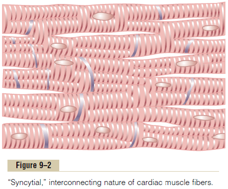
At each intercalated disc the cell membranes fuse with one another in such a way that they form perme-able “communicating” junctions (gap junctions) that allow almost totally free diffusion of ions. Therefore, from a functional point of view, ions move with ease in the intracellular fluid along the longitudinal axes of the cardiac muscle fibers, so that action potentials travel easily from one cardiac muscle cell to the next, past the intercalated discs. Thus, cardiac muscle is a syncytium of many heart muscle cells in which thecardiac cells are so interconnected that when one of these cells becomes excited, the action potential spreads to all of them, spreading from cell to cell throughout the latticework interconnections.
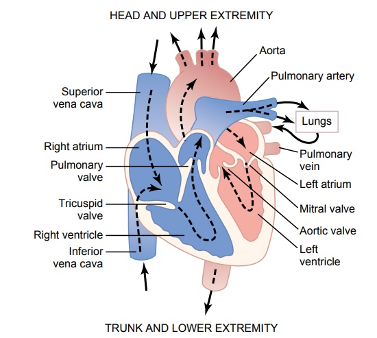
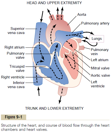
The heart actually is composed of two syncytiums: the atrial syncytium that constitutes the walls of the two atria, and the ventricular syncytium that consti-tutes the walls of the two ventricles. The atria are sepa-rated from the ventricles by fibrous tissue that surrounds the atrioventricular (A-V) valvular open-ings between the atria and ventricles. Normally, poten-tials are not conducted from the atrial syncytium into the ventricular syncytium directly through this fibrous tissue. Instead, they are conducted only by way of a specialized conductive system called the A-V bundle, a bundle of conductive fibers several millimeters in diameter.
This division of the muscle of the heart into two functional syncytiums allows the atria to contract a short time ahead of ventricular contraction, which is important for effectiveness of heart pumping.
Related Topics