Chapter: Medical Physiology: Secretory Functions of the Alimentary Tract
Physiologic Anatomy of Biliary Secretion
Physiologic Anatomy of Biliary Secretion
Bile is secreted in two stages by the liver: (1) The initial portion is secreted by the principal functional cells of the liver, thehepatocytes; this initial secretion contains large amounts of bile acids, cholesterol, and other organic constituents. It is secreted into minute bilecanaliculi that originate between the hepatic cells.
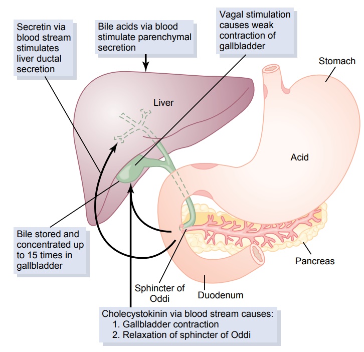
(2) Next, the bile flows in the canaliculi toward the interlobular septa, where the canaliculi empty into ter-minal bile ducts and then into progressively largerducts, finally reaching the hepatic duct and commonbile duct. From these the bile either empties directlyinto the duodenum or is diverted for minutes up to several hours through the cystic duct into the gall-bladder, shown in Figure 64–11.
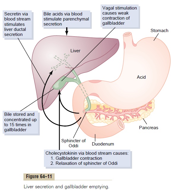
In its course through the bile ducts, a second portion of liver secretion is added to the initial bile. This addi-tional secretion is a watery solution of sodium and bicarbonate ions secreted by secretory epithelial cells that line the ductules and ducts. This second secretion sometimes increases the total quantity of bile by as much as an additional 100 per cent. The second secre-tion is stimulated especially by secretin, which causes release of additional quantities of bicarbonate ions to supplement the bicarbonate ions in pancreatic secre-tion (for neutralizing acid that empties into the duo-denum from the stomach).
Storing and Concentrating Bile in the Gallbladder. Bile issecreted continually by the liver cells, but most of it is normally stored in the gallbladder until needed in the duodenum. The maximum volume that the gallbladder can hold is only 30 to 60 milliliters. Nevertheless, as much as 12 hours of bile secretion (usually about 450 milliliters) can be stored in the gallbladder because water, sodium, chloride, and most other small elec-trolytes are continually absorbed through the gall-bladder mucosa, concentrating the remaining bile constituents that contain the bile salts, cholesterol, lecithin, and bilirubin.
Most of this gallbladder absorption is caused by active transport of sodium through the gallbladder epithelium, and this is followed by secondary absorp-tion of chloride ions, water, and most other diffusible constituents. Bile is normally concentrated in this way about 5-fold, but it can be concentrated up to a maximum of 20-fold.
Composition of Bile. Table 64–2 gives the composition ofbile when it is first secreted by the liver and then after it has been concentrated in the gallbladder. This table shows that by far the most abundant substances secreted in the bile are bile salts, which account for about one half of the total solutes also in the bile. Also secreted or excreted in large concentrations are biliru-bin, cholesterol, lecithin, and the usual electrolytes of plasma.
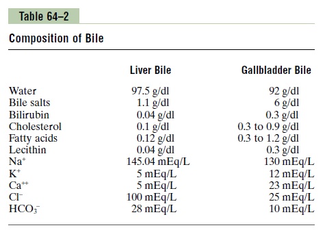
In the concentrating process in the gallbladder, water and large portions of the electrolytes (except calcium ions) are reabsorbed by the gallbladder mucosa; essentially all other constituents, especially the bile salts and the lipid substances cholesterol and lecithin, are not reabsorbed and, therefore, become highly concentrated in the gallbladder bile.
Emptying of the Gallbladder—Stimulatory Role of Cholecys-tokinin. When food begins to be digested in the uppergastrointestinal tract, the gallbladder begins to empty, especially when fatty foods reach the duodenum about 30 minutes after a meal. The mechanism of gallblad-der emptying is rhythmical contractions of the wall of the gallbladder, but effective emptying also requires simultaneous relaxation of the sphincter of Oddi, which guards the exit of the common bile duct into the duodenum.
By far the most potent stimulus for causing the gall-bladder contractions is the hormone cholecystokinin.
This is the same cholecystokinin discussed earlier that causes increased secretion of digestive enzymes by the acinar cells of the pancreas. The stimulus for cholecys-tokinin entry into the blood from the duodenal mucosa is mainly the presence of fatty foods in the duodenum.
In addition to cholecystokinin, the gallbladder is stimulated less strongly by acetylcholine-secreting nerve fibers from both the vagi and the intestinal enteric nervous system. They are the same nerves that promote motility and secretion in other parts of the upper gastrointestinal tract.
In summary, the gallbladder empties its store of con-centrated bile into the duodenum mainly in response to the cholecystokinin stimulus that itself is initiated mainly by fatty foods. When fat is not in the food, the gallbladder empties poorly, but when significant quantities of fat are present, the gallbladder normally empties completely in about 1 hour. Figure 64–11 sum-marizes the secretion of bile, its storage in the gall-bladder, and its ultimate release from the bladder to the duodenum.
Related Topics