Chapter: Human Nervous System and Sensory Organs : Functional Systems
Pathway of the Protopathic Sensibility
Pathway of the Protopathic Sensibility
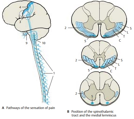
The thin, poorly myelinated or unmyelinated nerve fibers for the senses of pain andtemperature originate from the small neurons of the spinal ganglia (A1) (1st neuron). Their centripetal axons enter the spinal cord through the lateral part of the posterior root. They bifurcate in Lissauer’s tract and terminate in the dorsal border region of the substantia gelatinosa and in the poste-rior horn. The secondary fibers cross to the opposite side and ascend in the anterolateral funiculus as lateralspinothalamic tract (B2) (2nd neuron). Thetract does not form a discrete fiber bundle but consists of loosely arranged fibers that are mixed with fibers of other systems. The fibers entering at various root levels join ventromedially. Thus, the sacral fibers lie at the surface, and the cervical fibers that joined last lie in the inner part of the an-terolateral funiculus.
The
input of impulses is controlled by de-scending fibers that originate in the
central region, in the anterior lobe of the cerebel-lum, and in the reticular
formation. These fibers terminate in the substantia gelati-nosa, a relay
station in which the peripheral impulses are modulated by the excitatory or
inhibitory influences of higher centers. Numerous axo-axonal synapses, which
are typical for presynaptic inhibition, have been demonstrated in the
substantia gelatinosa.
In the
medulla oblongata, the lateral spinothalamic tract (spinal lemniscus) is lo-cated at its lateral margin above the olive
and gives off numerous collaterals to the re-ticular formation. Here, too, a
considerable portion of the fibers (spinoreticular
tract) terminate. The reticular formation is part of the ascending
activation system, the stimulation of which puts the organism into a state of
alertness. Hence, the impulses transmitted via the pain pathway not only cause a conscious sensation but also
in-crease the attention via the reticular forma-tion. By contrast, the pathway
of the epicritic sensibility runs through the brain stem without giving off any
collaterals.

The
spinothalamic fibers join the mediallemniscus
in the midbrain and take a dor-solateral position. A large portion of them
terminate on the cells of the ventral
poste-rior nucleus of thalamus (AC3)
(3rd neuron)in somatotopic organization, predomi-nantly in a ventral
parvocellular region. Ter-tiary fibers extend from here to the postcen-tral
region (A4). Other spinothalamic
fibers terminate in other thalamic nuclei, for ex-ample, in the intralaminar
nuclei.
The anterior spinothalamic tract (B5) transmits crude senses of touch and pressure.
Its fibers cross from the posterior horn (2nd neuron) to the contralateral
anterior funiculus. The position of the tract in the medulla oblongata is a
matter of controversy. It is though to lie either medi-ally to the medial
lemniscus (B6) or laterally to the
olive (B7). In the pons and
midbrain, the fibers join the medial lemniscus (B8) and terminate on the cells of the ventral pos-terior nucleus of thalamus (3rd neuron).
Pain and
temperature fibers for the face
andsinuses originate from the neurons of thetrigeminal ganglion (A9), the centripetal axons of which
terminate in the spinal nu-cleus of the
trigeminal nerve (AB10). In the
spinal tract of the trigeminal nerve, the pain-transmitting fibers are thought
to lie laterally and those transmit-ting temperature further medially. The
sec-ondary trigeminal fibers (B11) (trigeminallemniscus ) join the medial
lemniscus.
Related Topics