Chapter: Medical Physiology: The Eye: III. Central Neurophysiology of Vision
Organization and Function of the Visual Cortex
Organization and Function of the Visual Cortex
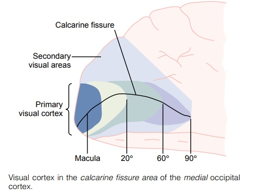
Figures 51–2 and 51–3 show the visual cortex located primarily on the medial aspect of the occipital lobes. Like the cortical representations of the other sensory systems, the visual cortex is divided into a primaryvisual cortex and secondary visual areas.
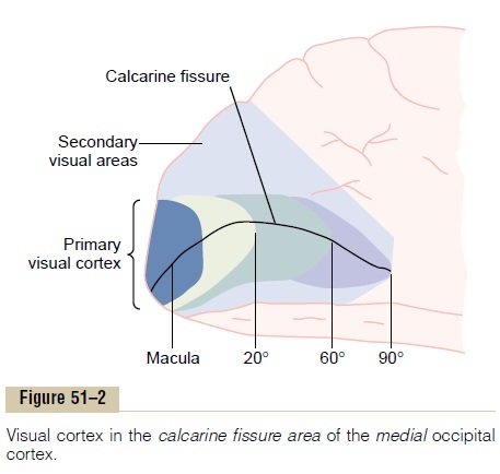
Primary Visual Cortex. The primary visual cortex (see Figure 51–2) lies in the calcarine fissure area, extend-ing forward from the occipital pole on the medial aspect of each occipital cortex. This area is the termi-nus of direct visual signals from the eyes. Signals from the macular area of the retina terminate near the occipital pole, as shown in Figure 51–2, while signals from the more peripheral retina terminate at or in con-centric half circles anterior to the pole but still along the calcarine fissure on the medial occipital lobe. The upper portion of the retina is represented superiorly and the lower portion inferiorly.
Note in the figure the especially large area that rep-resents the macula. It is to this region that the retinal fovea transmits its signals. The fovea is responsible for the highest degree of visual acuity. Based on retinal area, the fovea has several hundred times as much rep-resentation in the primary visual cortex as do the most peripheral portions of the retina.
The primary visual cortex is also called visual areaI. Still another name is the striate cortex because thisarea has a grossly striated appearance.
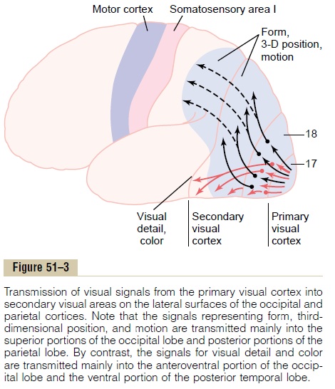
Secondary Visual Areas of the Cortex. The secondary visualareas, also called visual association areas, lie lateral, anterior, superior, and inferior to the primary visual cortex. Most of these areas also fold outward over the lateral surfaces of the occipital and parietal cortex, as shown in Figure 51–3. Secondary signals are transmit-ted to these areas for analysis of visual meanings. For instance, on all sides of the primary visual cortex is Brodmann’s area 18 (see Figure 51–3), which is wherevirtually all signals from the primary visual cortex pass next. Therefore, Brodmann’s area 18 is called visualarea II, or simply V-2. The other, more distant second-ary visual areas have specific designations—V-3, V-4, and so forth—up to more than a dozen areas. The importance of all these areas is that various aspects of the visual image are progressively dissected and analyzed.
Layered Structure of the Primary Visual Cortex
Like almost all other portions of the cerebral cortex, the primary visual cortex has six distinct layers, as shown in Figure 51–4. Also, as is true for the other sensory systems, the geniculocalcarine fibers terminate mainly in layer IV. But this layer, too, is organized into subdivisions. The rapidly conducted signals from the Y retinal ganglion cells terminate in layer IVca, and from there they are relayed vertically both outward toward the cortical surface and inward toward deeper levels.
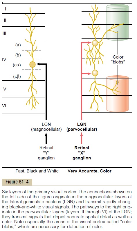
The visual signals from the medium-sized optic nerve fibers, derived from the X ganglion cells in the retina, also terminate in layer IV, but at points differ-ent from the Y signals. They terminate in layers IVa and IVcb, the shallowest and deepest portions of layer IV, shown to the right in Figure 51–4. From there, these signals are transmitted vertically both toward the surface of the cortex and to deeper layers. It is these X ganglion pathways that transmit the accurate point-to-point type of vision as well as color vision.
Vertical Neuronal Columns in the Visual Cortex. The visualcortex is organized structurally into several million vertical columns of neuronal cells, each column having a diameter of 30 to 50 micrometers. The same vertical columnar organization is found throughout the cere-bral cortex for the other senses as well (and also in the motor and analytical cortical regions). Each column represents a functional unit. One can roughly calculate that each of the visual vertical columns has perhaps 1000 or more neurons.
After the optic signals terminate in layer IV, they are further processed as they spread both outward and inward along each vertical column unit. This process-ing is believed to decipher separate bits of visual infor-mation at successive stations along the pathway. The signals that pass outward to layers I, II, and III even-tually transmit signals for short distances laterally in the cortex. Conversely, the signals that pass inward to layers V and VI excite neurons that transmit signals much greater distances.
“Color Blobs” in the Visual Cortex. Interspersed among theprimary visual columns as well as among the columns of some of the secondary visual areas are special column-like areas called color blobs. They receive lateral signals from adjacent visual columns and are activated specifically by color signals. Therefore, it is presumed that these blobs are the primary areas for deciphering color.
Interaction of Visual Signals from the Two Separate Eyes.
Recall that the visual signals from the two separate eyes are relayed through separate neuronal layers in the lateral geniculate nucleus. These signals still remain separated from each other when they arrive in layer IV of the primary visual cortex. In fact, layer IV is interlaced with stripes of neuronal columns, each stripe about 0.5 millimeter wide; the signals from one eye enter the columns of every other stripe, alter-nating with signals from the second eye. This cortical area deciphers whether the respective areas of the two visual images from the two separate eyes are “in register” with each other—that is, whether correspon-ding points from the two retinas fit with each other. In turn, the deciphered information is used to adjust the directional gaze of the separate eyes so that they will fuse with each other (be brought into “register”). The information observed about degree of register of images from the two eyes also allows a person to dis-tinguish the distance of objects by the mechanism of stereopsis.
Two Major Pathways for Analysis of Visual Information—(1) The Fast “Position” and “Motion” Pathway; (2) The Accurate Color Pathway
Figure 51–3 shows that after leaving the primary visual cortex, the visual information is analyzed in two major pathways in the secondary visual areas.
1. Analysis of Third-Dimensional Position, Gross Form, and Motion of Objects. One of the analytical pathways,demonstrated in Figure 51–3 by the black arrows, ana-lyzes the third-dimensional positions of visual objects in the space around the body. This pathway also ana-lyzes the gross physical form of the visual scene as well as motion in the scene. In other words, this pathway tells where every object is during each instant and whether it is moving. After leaving the primary visual cortex, the signals flow generally into the posteriormidtemporal area and upward into the broad occipi-toparietal cortex. At the anterior border of the parietalcortex, the signals overlap with signals from the pos-terior somatic association areas that analyze three-dimensional aspects of somatosensory signals. The signals transmitted in this position-form-motion pathway are mainly from the large Y optic nerve fibers of the retinal Y ganglion cells, transmitting rapid signals but depicting only black and white with no color.
2. Analysis of Visual Detail and Color. The red arrows in Figure 51–3, passing from the primary visual cortex into secondary visual areas of the inferior, ventral, and medial regions of the occipital and temporal cortex, show the principal pathway for analysis of visual detail. Separate portions of this pathway specifically dissect out color as well. Therefore, this pathway is concerned with such visual feats as recognizing letters, reading, determining the texture of surfaces, deter-mining detailed colors of objects, and deciphering from all this information what the object is and what it means.
Related Topics