Chapter: Human Nervous System and Sensory Organs : The Eye
Optic Nerve - Structure of the Eye
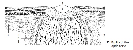
Optic Nerve
The
nerve fibers of the retina extend in bundles to the papilla of the optic nerve (D3)
where they unite to form the optic nerve before exiting the eyeball. Sclera and
choroidea are very thin at the site of pene-tration, and the sclera is
perforated (laminacribrosa) (D4). Once the extremely delicatenerve
fibers have passed the sclera, they be-come enveloped by myelin sheaths. The
optic nerve is actually a fiber tract of the CNS and contains astrocytes and
oligoden-drocytes; hence, its nerve fibers do not have Schwann cell sheaths. As
part of the brain, the optic nerve is surrounded by mengines. The dural sheath (D5) and the arachnoidsheath (D6) merge with the sclera (D7). Be-tween arachnoid sheath and pial
sheath (D8) lies a CSF-filled
space (D9) which makes a shift
between nerve and sheath possible. A number of septae from the pia mater extend
between the nerve bundles.

Related Topics