Chapter: Medical Physiology: The Chemical Senses-Taste and Smell
Olfactory Membrane - Sense of Smell
Olfactory Membrane
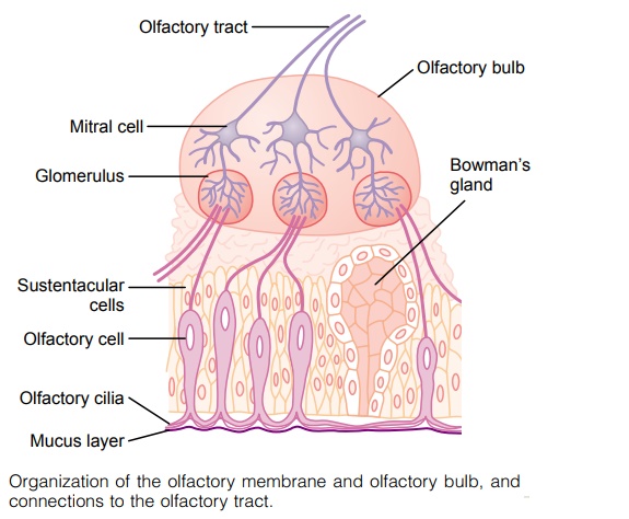
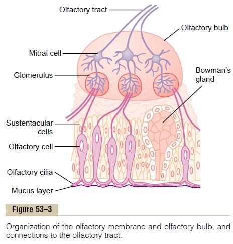
The olfactory membrane, the histology of which is shown in Figure 53–3, lies in the superior part of each nostril. Medially, the olfactory membrane folds down-ward along the surface of the superior septum; later-ally, it folds over the superior turbinate and even over a small portion of the upper surface of the middle turbinate. In each nostril, the olfactory membrane has a surface area of about 2.4 square centimeters.
Olfactory Cells. The receptor cells for the smell sensa-tion are the olfactory cells (see Figure 53–3), which are actually bipolar nerve cells derived originally from the central nervous system itself. There are about 100 million of these cells in the olfactory epithelium inter-spersed among sustentacular cells, as shown in Figure 53–3. The mucosal end of the olfactory cell forms a knob from which 4 to 25 olfactory hairs (also called olfactory cilia), measuring 0.3 micrometer in diameterand up to 200 micrometers in length, project into the mucus that coats the inner surface of the nasal cavity. These projecting olfactory cilia form a dense mat in the mucus, and it is these cilia that react to odors in the air and stimulate the olfactory cells, as discussed later. Spaced among the olfactory cells in the olfactory membrane are many small Bowman’s glands that secrete mucus onto the surface of the olfactory membrane.
Related Topics