Chapter: Ophthalmology: Retina
Normal and Abnormal Fundus Findings in General
Normal and Abnormal Fundus Findings in General
Normal fundus:
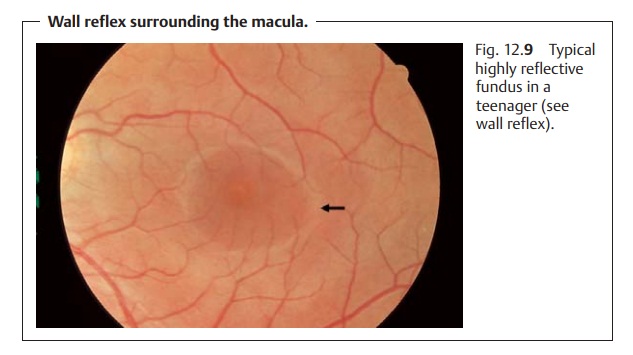
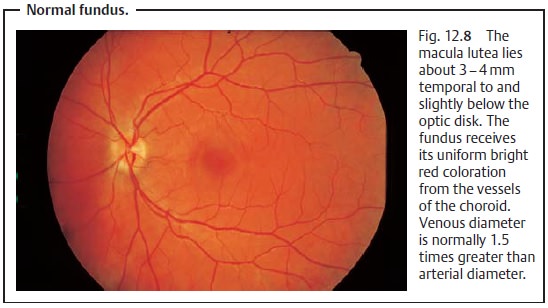
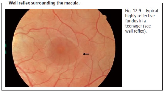
The retina is normally completely transparent without any intrinsic color. It receives its uniform bright red coloration from the vascula-ture of the choroid (Fig. 12.8). The vessels of the choroid themselves are obscured by the retinal pigment epithelium. Loss of transparency of the retina is a sign of an abnormal process (for example in retinal edemas, the retina appears whitish yellow). The optic disk is normally a sharply defined, yel-lowish orange structure (in teenagers it is pale pink, and in young children sig-nificantly paler) that may exhibit a central depression known as the optic or physiologic cup. Light reflection on the inner limiting membrane will nor-mally produce multiple light reflexes on the fundus. Teenagers will also exhibit a normal foveal reflex and wall reflex surrounding the macula, which is caused by the transition from the depression of the macular to the higher level of the retina (Fig. 12.9).
Age-related changes:
Theopticdisk turns
pale yellow with age, and oftenthe optic cup will become shallow and will be
surrounded by a region of choroidal atrophy. The fundus will become dull and nonreflective. Drusen will be visible in the retinal pigment epithelium and middle
peripheral reticu-lar proliferations of
pigment epithelium will be present. The
arterioles will be elongated due to loss of elasticity with irregular filling due to thickening of
thevascular walls. Meandering of the
venules will be present with crossing
signs, i.e., the sclerotic artery will be seen to compress the vein at the
arteriovenous crossing, reducing the diameter of the column of venous blood. In
extreme cases venous blood flow will be cut off completely.
Abnormal changes in the fundus:
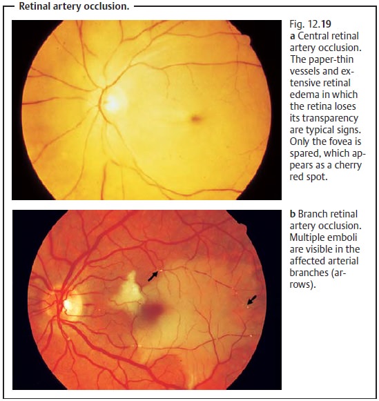
As a rule,loss of transparency of the retinais a
sign of an abnormal process. For example in a retinal edema, the retina appears
whitish yellow (see Fig. 12.19). A
distinctive feature of abnormal reti-nal and choroidal changes is that the type
and appearance of these changes permit precise topographic localization of the respective abnormal process when
the diagnosis is made. The ophthalmoscopic image will usually allow one to
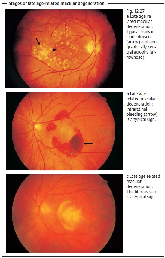
determine in which of the layers shown in Fig. 12.2 the process is occurring. For example, in Fig. 12.27 (nonexudative age-related macular
degeneration) one may see that the drusen and atrophy are located in the
ret-inal pigment epithelium; the structures above it are not affected, as is
apparent from the intact vascular structures.
Related Topics