Chapter: 11th Zoology : Chapter 10 : Neural Control and Coordination
Neuron as a structural and functional unit of Neural system
Neuron as a structural and functional unit of Neural
system
A neuron is a microscopic structure composed of
three major parts namely cell body
(soma), dendrites and axon. The cell body is the spherical
part of the neuron that contains all the cellular organelles as a typical cell
(except centriole). The plasma
membrane covering the neuron is called neurilemma
and the axon is axolemma. The
repeatedly branched short fibres coming out of the cell body are called dendrites, which transmit impulses
towards the cell body. The cell body and the dendrites contain cytoplasm and
granulated endoplasmic reticulum called Nissl’s
granules.
An axon is a long fibre that arises from a cone
shaped area of the cell body called the Axon
hillock and ends at the branched distal end. Axon hillock is the place
where the nerve impulse is generated
in the motor neurons. The axon of one-neuron branches and forms connections
with many other neurons. An axon contains the same organelles found in the
dendrites and cell body but lacks Nissl’s granules and Golgi apparatus.
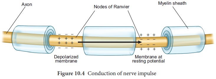
The axon, particularly of peripheral nerves is surrounded by Schwann cells (a type of glial cell) to form myelin sheath, which act as an insulator. Myelin sheath is associated only with the axon; dendrites are always non-myelinated. Schwann cells are not continuous along the axon; so there are gaps in the myelin sheath between adjacent Schwann cells. These gaps are called Nodes of Ranvier . Large myelinated nerve fibres conduct impulses rapidly, whereas non-myelinated fibres conduct impulses quite slowly (Figure 10.1).
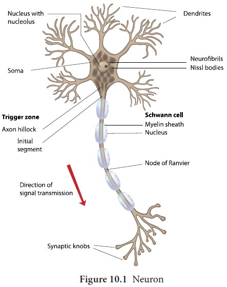
Each branch at the distal end of the axon
terminates into a bulb like structure called synaptic knob which possesses
synaptic vesicles filled with
neurotransmitters. The axon
transmits nerve impulses away from the cell body to an inter neural space or to a neuro-muscular
junction.
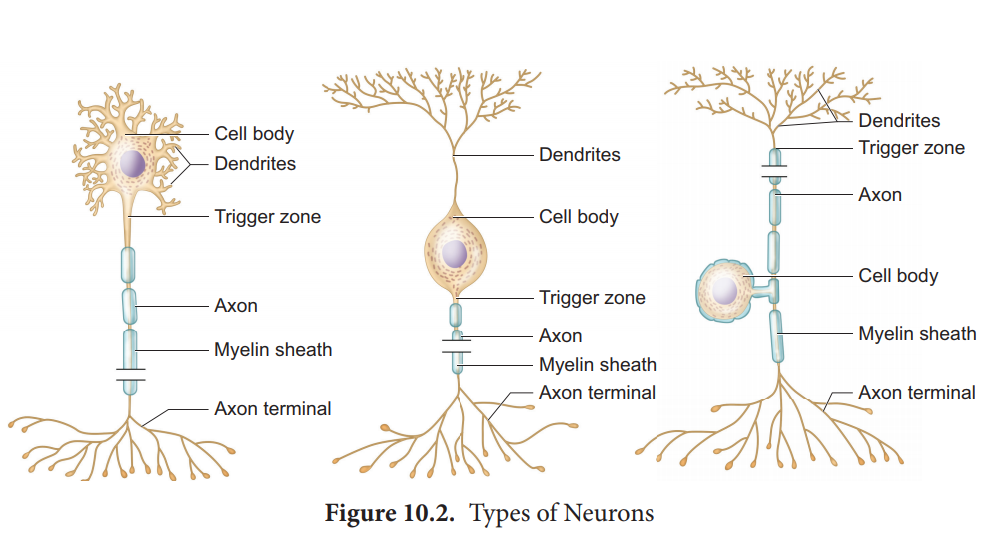
The neurons are divided into three types based on
number of axon and dendrites they possess (Figure.10.2).
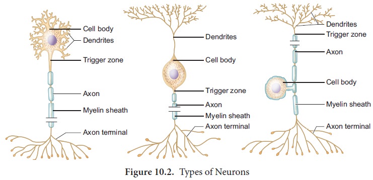
1.
Multipolar
neurons have many processes with one axon and two or more dendrites. They are mostly interneurons.
2.
Bipolar
neurons have two processes with one axon and one dendrite. These are found in the retina of the eye, inner ear and the
olfactory area of the brain.
3.
Unipolar
neurons have a single short process and one axon. Unipolar neurons are located in the ganglia of cranial and spinal nerves.
1. Generation and conduction of nerve impulses
This section deals with how the nerve impulses are produced and conducted in our body. Sensation felt in the sensory organs are carried by the nerve fibres in the form of electrical impulses. A nerve impulse is a series of electrical impulses, which travel along the nerve fibre. Inner to the axolemma, the cytoplasm contains the intracellular fluid (ICF ) with large amounts of potassium and magnesium phosphate along with negatively charged proteins and other organic molecules.The extra cellular fluid (ECF) found outside the axolemma contains large amounts of sodium chloride, bicarbonates, nutrients and oxygen for the cell; and carbon dioxide and metabolic wastes released by the neuronal cells. The ECF and ICF (cytosol) contains negatively charged particles (anions) and positively charged particles (cations) . These charged particles are involved in the conduction of impulses.
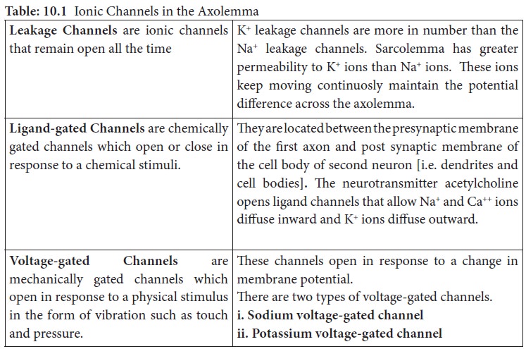
The neurons maintain an uneven distribution of
various inorganic ions across their axolemma for transmission of impulses. This
unequal distribution of ions establishes the membrane potential across the
axolemma. The axolemma contains a variety of membrane proteins that act as
ionic channels and regulates the movement of ions across the axolemma. (Shown
in Table 10.1).
2. Transmission of impulses
The transmission of impulse involves two main
phases; Resting membrane potential
and Action membrane potential.
Resting
membrane Potential: The electrical potential difference across
theplasma membrane of a resting
neuron is called the resting potential during which the interior of the cell is
negative due to greater efflux of K+ outside the cell than Na+ influx into the
cell. When the axon is not conducting any impulses i.e. in resting condition,
the axon membrane is more
The
axoplasm contains high concentration of K+ and negatively charged proteins and
low concentration of Na+ ions.
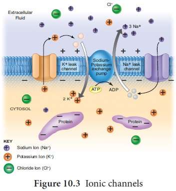
In contrast, fluid outside the axon (ECF) contains
low concentration of K+ and high concentration of Na+, and this forms a
concentration gradient. This ionic gradient across the resting membrane is
maintained by ATP driven Sodium-Potassium pump,
In this state, the cell membrane is said to be polarized. In neuron, the resting
membrane potential ranges from - 40mV
to -90mV, and its normal value
is -70mV. The minus sign indicates
that the inside of the cell is
negative with respect to the outside (Figure 10.4).
Action membrane potential
An action potential occurs when a neuron sends information
down an axon, away from the cell body. It includes following phases,
depolarization, repolarisation and hypo polarization.
Depolarization
– Reversal of polarity When a nerve fibre is stimulated, sodium voltage-gate opens and makes
the axolemma permeable to Na+ ions; meanwhile the potassium voltage gate closes. As a result, the rate of flow
of Na+ ions into the axoplasm exceeds the rate of flow of
K+ ions to the outside fluid [ECF].
Therefore, the axolemma becomes
positively charged inside and negatively charged outside. This reversal of
electrical charge is called Depolarization.
During depolarization, when enough Na+ ions enter
the cell, the action potential reaches a certain level, called threshold potential [-55mV]. The
particular stimulus which is able to bring the membrane potential to threshold
is called threshold stimulus.
The action potential occurs in response to a threshold stimulus but does not occur at subthreshold stimuli. This is called all or none principle. Due to the rapid influx of Na+ ions, the membrane potential shoots rapidly
up to +45mV which is called the Spike
potential.
Repolarisation [Falling Phase]
When the membrane reaches the spike potential, the sodium voltage-gate closes and potassium voltage-gate opens. It checks
influx of Na+ ions and
initiates the efflux of K+ions which
lowers the number of positive ions within the cell. Thus, the potential falls
back towards the resting potential. The reversal of membrane potential inside
the axolemma to negative occurs due to the efflux of K+ ions. This
is called Repolarisation.
Hyperpolarization
If repolarization becomes more negative than the
resting potential -70 mV to about - 90 mV, it is called Hyperpolarization. During this, K+ ion gates are more
permeable to K+ even after reaching the threshold level as it closes
slowly; hence called Lazy gates. The membrane potential returns
to its original resting state when K+ ion channels close completely. During hyperpolarization the Na+
voltage gate -remains closed (Figure 10.5).

Conduction Speed of a nerve impulse
The conduction speed of a nerve impulse depends on
the diameter of axon. The greater the axon’s diameter, the faster is the
conduction. The myelinated axon
conducts the impulse faster than the non-myelinated axon. The voltage-gated Na+
and K+ channels are concentrated at the nodes
of Ranvier. As a result, the impulse jumps node to node, rather than
travelling the entire length of the nerve fibre. This mechanism of conduction
is called Saltatory Conduction.
Nerve impulses travel at the speed of 1-300 m/s.
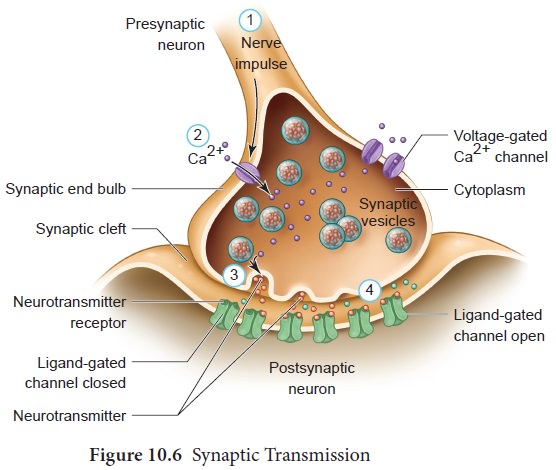
3. Synaptic transmission
The junction between two neurons is called a Synapse through which a nerve
The first neuron involved
in the synapse forms the pre-synaptic neuron and the second neuron is the
post-synaptic neuron. A small gap between the pre and postsynaptic membranes is
called Synaptic Cleft that forms a structural gap and a functional bridge
between neurons. The axon terminals contain synaptic vesicles filled with
neurotransmitters. When an impulse [action potential] arrives at the axon
terminals, it depolarizes the pre-synaptic membrane, opening the voltage gated
calcium channels. Influx of calcium ions stimulates the synaptic vesicles
towards the pre-synaptic membrane and fuses with it. In the neurilemma, the
vesicles release their neurotransmitters into the synaptic cleft by exocytosis.
Thereleased neurotransmitters bind to their specific receptors on the
post-synaptic membrane, responding to chemical signals. The entry of the ions
can generate new potential in the post-synaptic neuron, which may be either
excitatory or inhibitory. Excitatory post-synaptic potential causes
depolarization whereas inhibitory post-synaptic potential causes
hyperpolarization of post-synaptic membrane (Figure 10.6).
Related Topics