Chapter: Essentials of Anatomy and Physiology: The Nervous System
Nerve Tissue
NERVE TISSUE
Nerve tissue was briefly described, so we will begin by reviewing what you already know and then add to it.
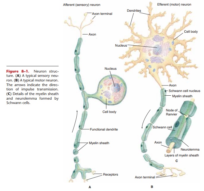
Nerve cells are called neurons, or nerve fibers. Whatever their specific functions, all neurons have the same physical parts. The cell body contains the nucleus (Fig. 8–1) and is essential for the continued life of the neuron. As you will see, neuron cell bodies are found in the central nervous system or close to it in the trunk of the body. In these locations, cell bodies are protected by bone. There are no cell bodies in the arms and legs, which are much more subject to injury.
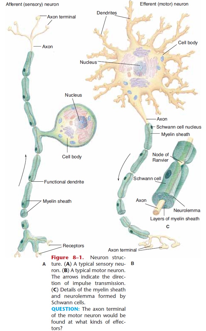
Figure 8–1. Neuron structure. (A) A typical sensory neu-ron. (B) A typical motor neuron. The arrows indicate the direc-tion of impulse transmission. (C) Details of the myelin sheath and neurolemma formed by Schwann cells.
QUESTION: The axon terminal of the motor neuron would be
Dendrites are processes (extensions) that transmit impulses toward the cell body. The one axon of a neu-ron transmits impulses away from the cell body. It is the cell membrane of the dendrites, cell body, and axon that carries the electrical nerve impulse.
In the peripheral nervous system, axons and den-drites are “wrapped” in specialized cells called Schwann cells (see Fig. 8–1). During embryonic development, Schwann cells grow to surround the neuron processes, enclosing them in several layers of Schwann cell membrane. These layers are the myelin sheath; myelin is a phospholipid that electrically insu-lates neurons from one another. Without the myelin sheath, neurons would short-circuit, just as electrical wires would if they were not insulated.
The spaces between adjacent Schwann cells, or seg-ments of the myelin sheath, are called nodes of Ranvier (neurofibril nodes). These nodes are the parts of the neuron cell membrane that depolarize when an electrical impulse is transmitted (see “The Nerve Impulse”).
The nuclei and cytoplasm of the Schwann cells are wrapped around the outside of the myelin sheath and are called the neurolemma, which becomes very important if nerves are damaged. If a peripheral nerve is severed and reattached precisely by microsurgery, the axons and dendrites may regenerate through the tunnels formed by the neurolemmas. The Schwann cells are also believed to produce a chemical growth factor that stimulates regeneration. Although this re-generation may take months, the nerves may eventu-ally reestablish their proper connections, and the person may regain some sensation and movement in the once-severed limb.
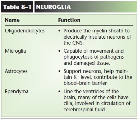
In the central nervous system, the myelin sheaths are formed by oligodendrocytes, one of the neu-roglia (glial cells), the specialized cells found only in the brain and spinal cord. Because no Schwann cells are present, however, there is no neurolemma, and regeneration of neurons does not occur. This is why severing of the spinal cord, for example, results in per-manent loss of function. Another kind of neuroglia are microglia,which are constantly moving, phagocytiz-ing cellular debris, damaged cells, and pathogens
Yet another type of glial cell is the astrocyte (liter-ally, “star cell”). In the embryo, these cells provide a framework for the migrating neurons that will form the brain. Thereafter, the extensions of astrocytes are wrapped around brain capillaries and contribute to theblood–brain barrier, which prevents potentially harmful waste products in the blood from diffusing out into brain tissue. These waste products are normal in the blood and tissue fluid, but brain tissue is much more sensitive to even low levels of them than are other tissues such as muscle tissue or connective tis-sue. The capillaries of the brain also contribute to this barrier, because they are less permeable than are other capillaries. A disadvantage of the blood–brain barrier is that some useful medications cannot cross it, and the antibodies produced by lymphocytes cross only with difficulty. This becomes an important considera-tion when treating brain infections or other diseases or disorders (Table 8–1 summarizes the functions of the neuroglia).
SYNAPSES
Neurons that transmit impulses to other neurons do not actually touch one another. The small gap or space between the axon of one neuron and the dendrites or cell body of the next neuron is called the synapse. Within the synaptic knob (terminal end) of the presy naptic axon is a chemical neurotransmitter that is released into the synapse by the arrival of an electrical nerve impulse (Fig. 8–2). The neurotransmitter dif-fuses across the synapse, combines with specific recep-tor sites on the cell membrane of the postsynaptic neuron, and there generates an electrical impulse that is, in turn, carried by this neuron’s axon to the next synapse, and so forth. A chemical inactivator at the cell body or dendrite of the postsynaptic neuron quickly inactivates the neurotransmitter. This pre-vents unwanted, continuous impulses, unless a new impulse from the first neuron releases more neuro-transmitter.
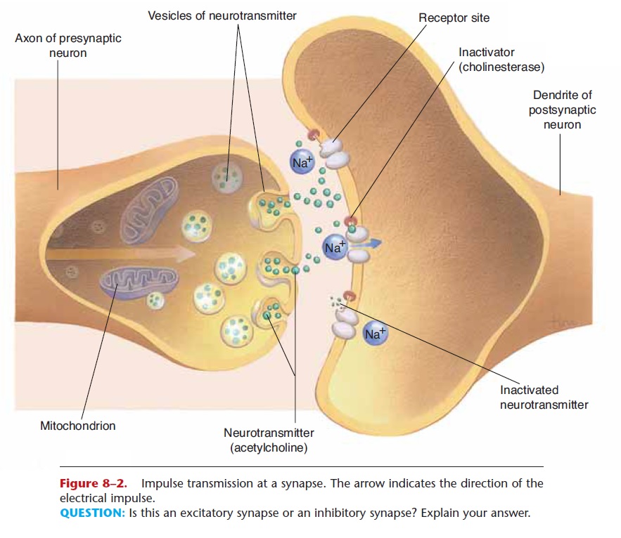
QUESTION: Is this an excitatory synapse or an inhibitory synapse? Explain your answer.
Many synapses are termed excitatory, because the neurotransmitter causes the postsynaptic neuron to depolarize (become more negative outside as Na+ ions enter the cell) and transmit an electrical impulse to another neuron, muscle cell, or gland. Some synapses, however, are inhibitory, meaning that the neurotrans-mitter causes the postsynaptic neuron to hyperpolar-ize (become even more positive outside as K+ ions leave the cell or Cl2 ions enter the cell) and therefore not transmit an electrical impulse. Such inhibitory synapses are important, for example, for slowing the heart rate, and for balancing the excitatory impulses transmitted to skeletal muscles. With respect to the skeletal muscles, this inhibition prevents excessive contraction and is important for coordination.
One important consequence of the presence of synapses is that they ensure one-way transmission of impulses in a living person. A nerve impulse cannot go backward across a synapse because there is no neuro-transmitter released by the dendrites or cell body. Neurotransmitters can be released only by a neuron’s axon, which does not have receptor sites for it, as does the postsynaptic membrane. Keep this in mind when we discuss the types of neurons later.
An example of a neurotransmitter is acetylcholine, which is found at neuromuscular junctions, in the CNS, and in much of the peripheral nervous system. Acetylcholine usually makes a postsynaptic membrane more permeable to Na+ ions, which brings about depolarization of the postsynaptic neuron. Cholin esterase is the inactivator of acetylcholine. There are many other neurotransmitters, especially in the cen-tral nervous system. These include dopamine, GABA, norepinephrine, glutamate, and serotonin. Each of these neurotransmitters has its own chemical inactiva-tor. Some neurotransmitters are reabsorbed into the neurons that secreted them; this process is called reuptake and also terminates the effect of the trans-mitter.
The complexity and variety of synapses make them frequent targets of medications. For example, drugs that alter mood or behavior often act on specific neu-rotransmitters in the brain, and antihypertensive drugs affect synapse transmission at the smooth mus-cle of blood vessels.
Related Topics