Chapter: Essentials of Anatomy and Physiology: Muscular System
Muscles of the head and Neck
Muscles of the head and Neck
The muscles of the head and neck include those involved in forming facial expressions, chewing, moving the tongue, swal-lowing, producing sounds, moving the eyes, and moving the head and neck.
Facial Expression
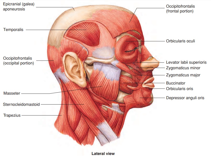
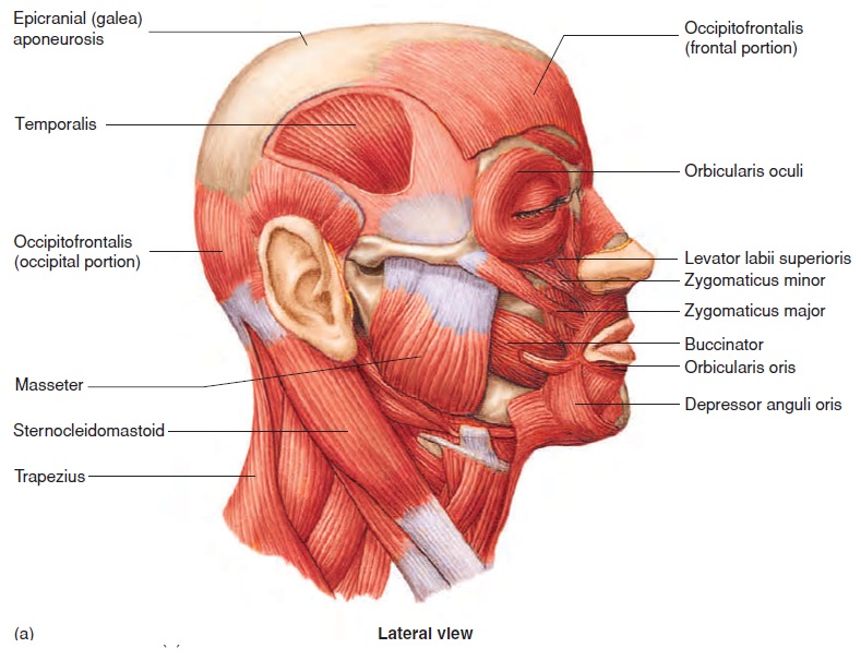
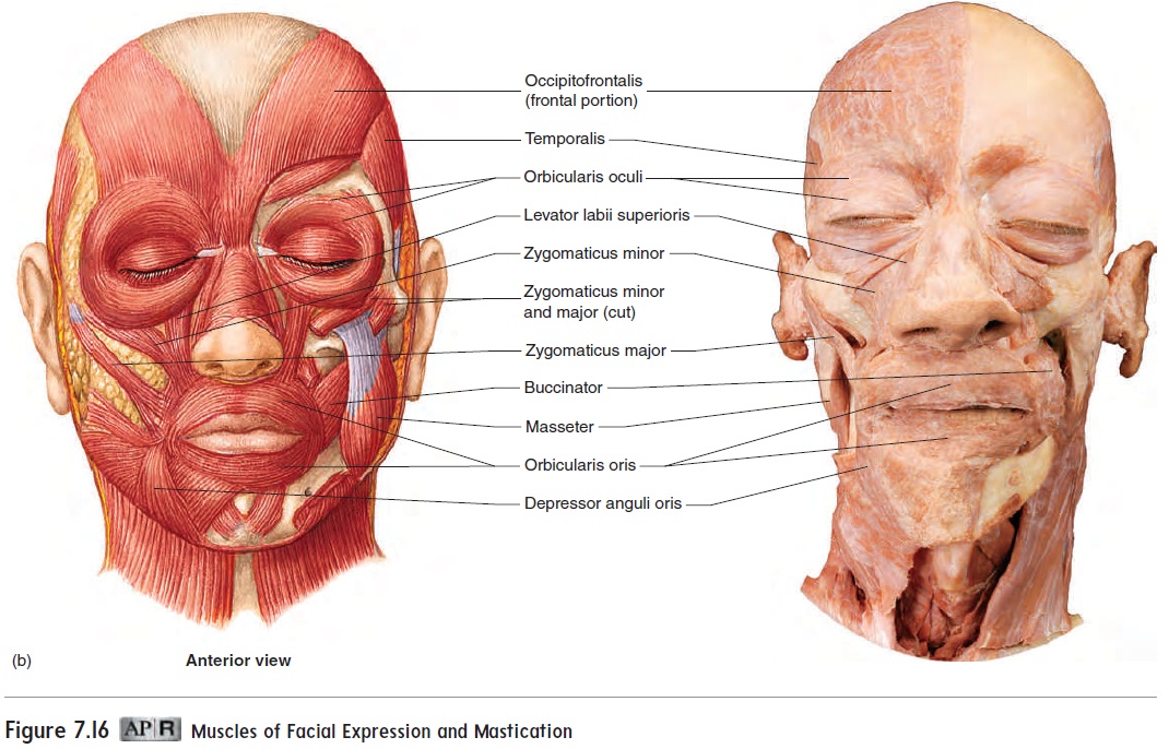
Several muscles act on the skin around the eyes and eyebrows (figure 7.16 and table 7.4). The occipitofrontalis (ok-sip′i-tō-frŭn-tā′lis) raises the eyebrows. The occipital and frontal portions of the muscle are connected by the epicranial aponeurosis. The orbicularis oculi (ōr-bik′ū-la′ris, circular+ok′ū-lı̄, eye) encirclethe eyes, tightly close the eyelids, and cause “crow’s feet” wrin-kles in the skin at the lateral corners of the eyes.
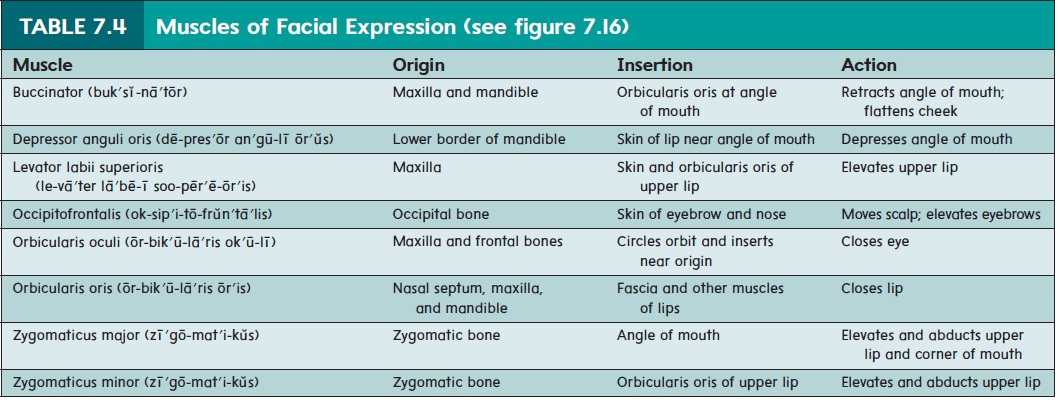
Several other muscles function in moving the lips and the skin surrounding the mouth (figure 7.16). The orbicularis oris (ōr′is; mouth), which encircles the mouth, and the buccinator (buk′sı̆-nā′tōr; bucca,cheek) are sometimes called the kissing muscles because they pucker the mouth. The buccinator also flattens the cheeks as in whistling or blowing a trumpet and is therefore sometimes called the trumpeter’s muscle. Smiling is accomplished primarily by the zygomaticus (zı̄′gō-mat′i-kŭs) muscles, which elevate the upper lip and corner of the mouth. Sneering is accomplished by the levator labii superioris (le-vā′ter lā′bē-ı̄ soo-pēr ′ē-ōr′is) because the muscle elevates one side of the upper lip. Frowning and pouting are largely per-formed by the depressor anguli oris (dē-pres′ŏr an′gū-lı̄ ōr′ŭs), which depresses the corner of the mouth.
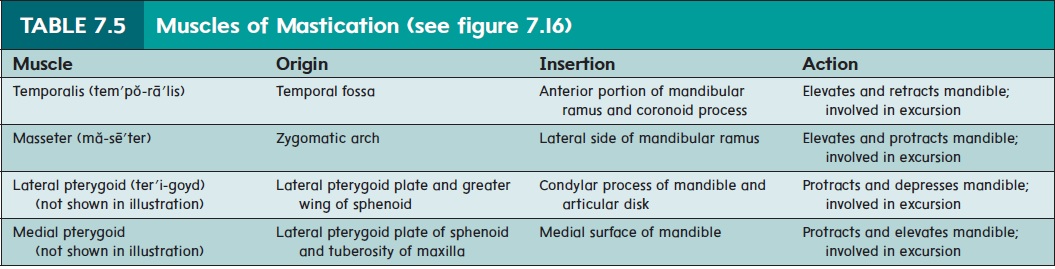
Mastication
The four pairs of muscles for chewing, or mastication (mas-ti-kā′shŭn), are some of the strongest muscles in the body (table 7.5). The temporalis (tem′ pŏ-rā′ lis) and masseter (ma-sē′ ter) muscles (figure 7.16) can be easily seen and felt on the side of the head during mastication. The pterygoid (ter′ ı̆-goyd) muscles, consist-ing of two pairs, are deep to the mandible
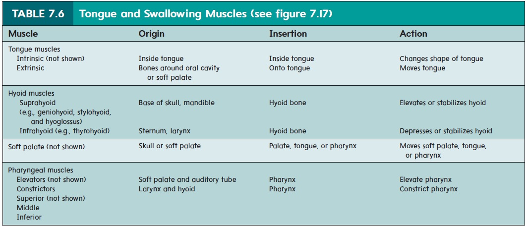
Tongue and Swallowing Muscles
The tongue is very important in mastication and speech. It moves food around in the mouth and, with the buccinator muscle, holds the food in place while the teeth grind the food. The tongue pushes food up to the palate and back toward the pharynx to initiate swal-lowing. The tongue consists of a mass of intrinsic muscles, which are located entirely within the tongue and change its shape. The extrinsic musclesare located outside the tongue but are attachedto and move the tongue (figure 7.17 and table 7.6).
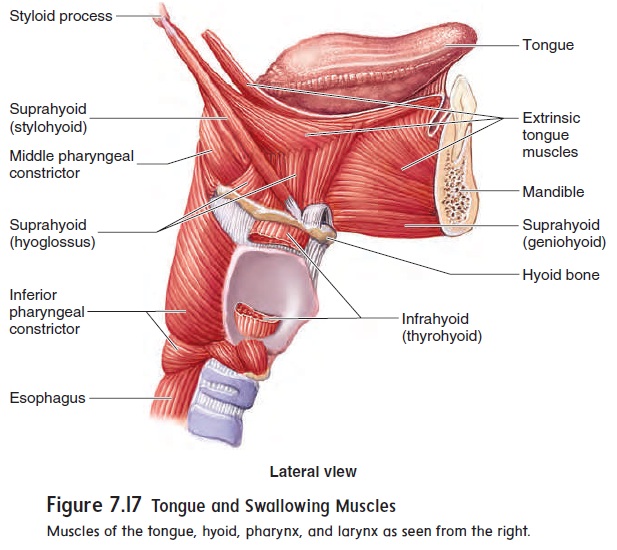
Swallowing involves a number of structures and their associ-ated muscles, including the hyoid muscles, soft palate, pharynx (throat), and larynx (voicebox). The hyoid (hi′oyd) muscles are divided into a suprahyoid group (superior to the hyoid bone) and an infrahyoid group (inferior to the hyoid bone) (figure 7.17 and table 7.6). When the suprahyoid muscles hold the hyoid bone in place from above, the infrahyoid muscles can elevate the larynx.
To observe this effect, place your hand on your larynx (Adam’s apple) and swallow.
The muscles of the soft palate close the posterior opening to the nasal cavity during swallowing, preventing food and liq-uid from entering the nasal cavity. When we swallow, muscles elevate the pharynx and larynx and then constrict the pharynx. Specifically, the pharyngeal (fă-rin′jē-ăl) elevators elevate the pharynx, and the pharyngeal constrictors constrict the pharynx from superior to inferior, forcing the food into the esophagus. Pharyngeal muscles also open the auditory tube, which connects the middle ear to the pharynx. Opening the auditory tube equal-izes the pressure between the middle ear and the atmosphere. This is why it is sometimes helpful to chew gum or swallow when ascending or descending a mountain in a car or changing altitude in an airplane
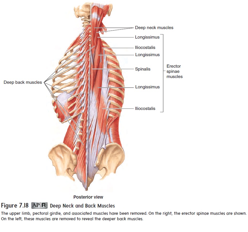
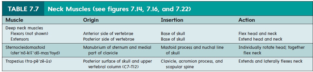
Neck Muscles
The deep neck muscles (figure 7.18 and table 7.7) include neck flexors, located along the anterior surfaces of the vertebral bodies, and neck extensors, located posteriorly. Rotation and lateral flexion of the head are accomplished by lateral and posterior neck muscles. The sternocleidomastoid (ster′nō-klı̄′dō-mas′toyd) muscle (see figure 7.16a), the prime mover of the lateral muscle group, is easi-ly seen on the anterior and lateral sides of the neck. Contraction of only one sternocleidomastoid muscle rotates the head. Contraction of both sternocleidomastoids flexes the neck or extends the head, depending on what the other neck muscles are doing. Torticollis (tōr′ti-kol′is; a twisted neck), or wry neck, may result from injury to one of the sternocleidomastoid muscles. It is sometimes caused by damage to a baby’s neck muscles during a difficult birth and usually can be corrected by exercising the muscle.
Related Topics