Chapter: Essentials of Anatomy and Physiology: The Nervous System
Meninges and Cerebrospinal Fluid
MENINGES AND CEREBROSPINAL FLUID
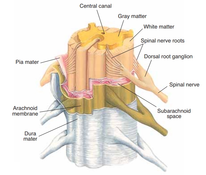
The connective tissue membranes that cover the brain and spinal cord are called meninges; the three layers are illustrated in Fig. 8–9. The thick outermost layer, made of fibrous connective tissue, is the dura mater (Latin for “tough mother”), which lines the skull and vertebral canal. The middle arachnoid membrane (arachnids are spiders) is made of web-like strands of connective tissue. The innermost pia mater (Latin for “gentle mother”) is a very thin membrane on the sur-face of the spinal cord and brain. Between the arach-noid and the pia mater is the subarachnoid space, which contains cerebrospinal fluid (CSF), the tissue fluid of the central nervous system.
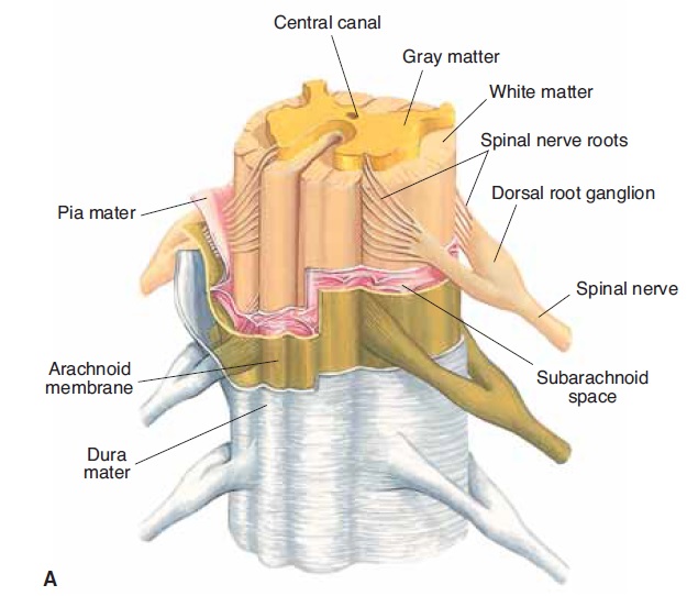
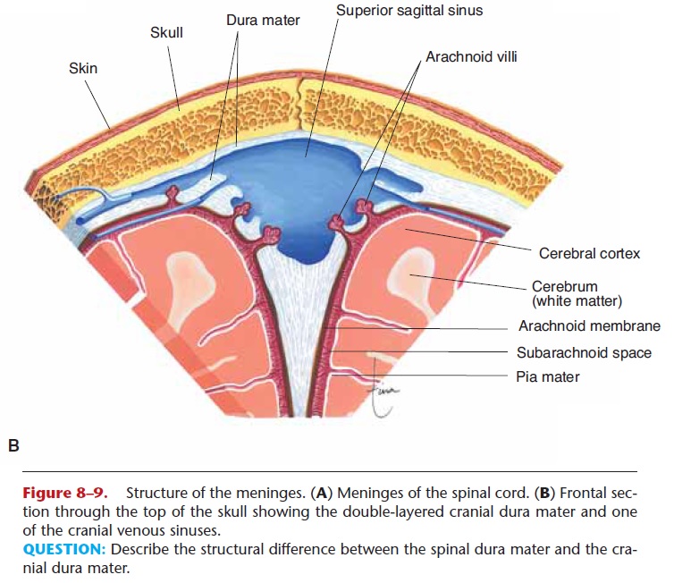
Figure 8–9. Structure of the meninges. (A) Meninges of the spinal cord. (B) Frontal sec-tion through the top of the skull showing the double-layered cranial dura mater and one of the cranial venous sinuses.
QUESTION: Describe the structural difference between the spinal dura mater and the cra-nial dura mater.
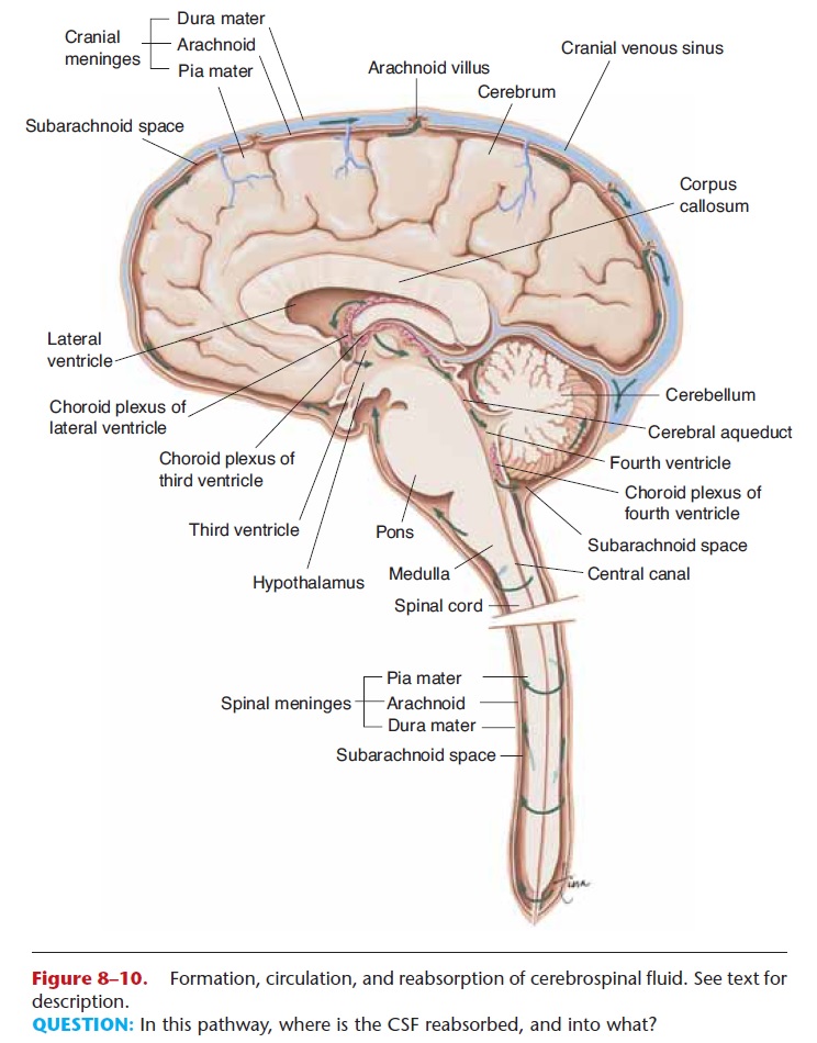
Recall the ventricles (cavities) of the brain: two lat-eral ventricles, the third ventricle, and the fourth ven-tricle. Each contains a choroid plexus, a capillary network that forms cerebrospinal fluid from blood plasma. This is a continuous process, and the cere-brospinal fluid then circulates in and around the cen-tral nervous system (Fig. 8–10).
From the lateral and third ventricles, cerebrospinal fluid flows through the fourth ventricle, then to the central canal of the spinal cord, and to the cranial and spinal subarachnoid spaces. As more cerebrospinal fluid is formed, you might expect that some must be reabsorbed, and that is just what happens. From the cranial subarachnoid space, cerebrospinal fluid is reab-sorbed through arachnoid villi into the blood in cranial venous sinuses (large veins within the double-layered cranial dura mater). The cerebrospinal fluid becomes blood plasma again, and the rate of reabsorption normally equals the rate of production.
Since cerebrospinal fluid is tissue fluid, one of its functions is to bring nutrients to CNS neurons and to remove waste products to the blood as the fluid is reabsorbed. The other function of cerebrospinal fluid is to act as a cushion for the central nervous system. The brain and spinal cord are enclosed in fluid-filled membranes that absorb shock. You can, for example, shake your head vigorously without harming your brain. Naturally, this protection has limits; very sharp or heavy blows to the skull will indeed cause damage to the brain.
Examination of cerebrospinal fluid may be used in the diagnosis of certain diseases.
Related Topics