Chapter: Essentials of Anatomy and Physiology: Nervous System
Meninges - Nervous System
Meninges
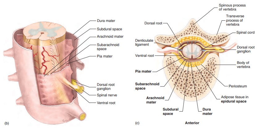
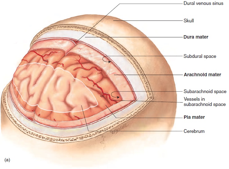
Three connective tissue membranes, the meninges (mĕ-nin′ jēz; meninx, membrane) (figure 8.34), surround and protect the brainand spinal cord. The most superficial and thickest of the meninges is the dura mater (doo′ ră mā′ ter; tough mother).
The dura mater around the brain consists of two layers, which function as a single layer but are physically separated into several regions to form dural folds and dural venous sinuses. Folds of dura mater extend into the longitudinal fissure between the two cerebral hemispheres and between the cerebrum and the cerebellum. The folds help hold the brain in place within the skull. The dural venous sinuses collect blood from the small veins of the brain and empty into the internal jugular veins, which exit the skull.
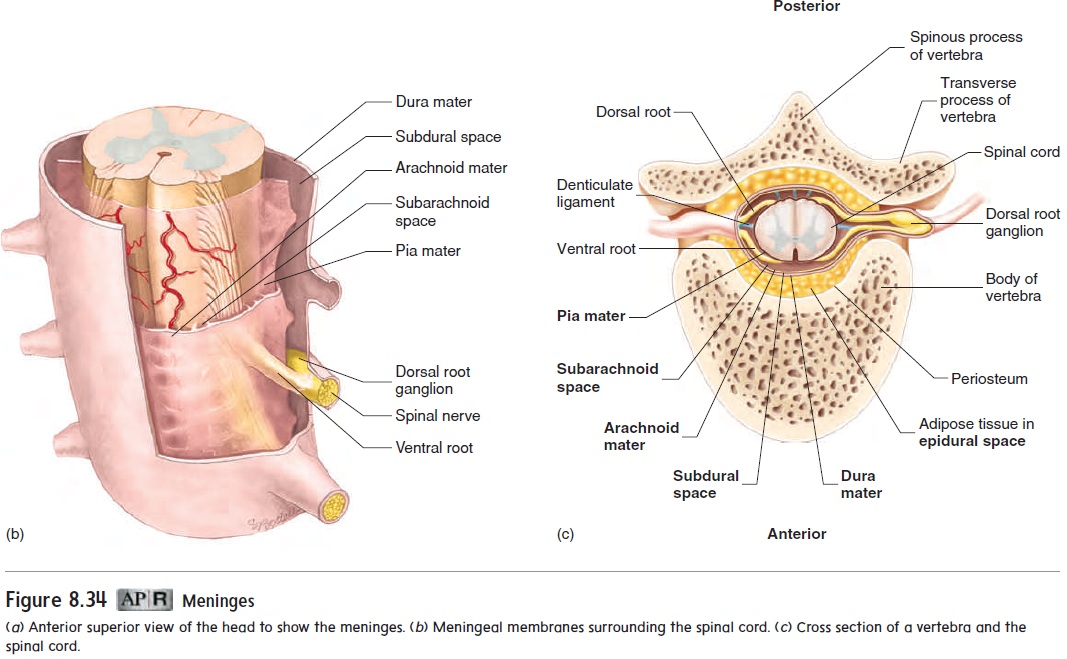
Within the skull, the dura mater adheres tightly to the cranial bones. In contrast, within the vertebral canal is an epidural space between the dura mater and the vertebrae (figure 8.34c). The epidural space is clinically important as the injection site for epidural anesthesia of the spinal nerves, which is often given towomen during childbirth.
The second meningeal membrane is the very thin, wispy arachnoid (ă-rak′noyd; spiderlike, as in cobwebs) mater. Thespace between the dura mater and the arachnoid mater is the sub-dural space, which is normally only a potential space containinga very small amount of serous fluid.
The spinal cord extends only to approximately the level of the second lumbar vertebra. Spinal nerves surrounded by meninges extend to the end of the vertebral column. Because there is no spinal cord in the inferior portion of the vertebral canal, a needlecan be introduced into the subarachnoid space at that level without damaging the spinal cord. Health professionals use such a needle to inject anesthetic into the area as a spinal block or to take a sample of cerebrospinal fluid in a spinal tap. The cerebrospinal fluid can then be examined for infectious agents (meningitis) or for blood (hemorrhage).
The third meningeal membrane, the pia mater (pı̄′ ă, pē′ ă; affectionate mother), is very tightly bound to the surface of the brain and spinal cord. Between the arachnoid mater and the pia mater is thesubarachnoid space, which is filled with cerebrospinal fluid and contains blood vessels.
Related Topics