Chapter: Obstetric and Gynecological Nursing : Abnormal Labour
Malpresentation and Malpostion
Malpresentation and Malpostion
Mal-presentation - A presentation other than
vertex Eg. Shoulder, face, brow and breech
Mal-position and mal-presentations have ill fitting presenting parts
compared to a well flexed vertex presentations in a normal pelvis.
Causes: - polyhydraminous
·
Abnormality of pelvis
·
Abnormal shape of uterus
·
Laxcity of uterine muscles
·
Multiple pregnancy
All ill fitting part is associated with (results in):
·
Early rupture of membrane with risk of cord prolapsed
·
Premature labour
·
Slow, irregular, short-lived contractions
·
Uncoordinated and excessively painful labour after rupture of membranes
·
Prolonged and obstructed labour
·
Post partum hemorrhage
·
Fetal and maternal distress
1. Breech Presentation
Definition: When the fetus lies with his
buttock in thelower pole of the uterus.
It occurs in 1: 40 cases of labor after 34 weeks
·
Breech with extended legs or frank breech- in this type of breech the
thighs are flexed and the legs are extended along the fetal abdomen. It is the
common one.
·
Complete breech the fetus lies in a flexed attitude and the legs are
flexed on the abdomen. The presenting part is bulky and consists of buttocks
external genitalia and both feet.
· Footling- one or both feet present because neither hips nor knees are fully flexed.
·
Knee presentation. On this case both the hips are extended with the
knees flexed.
Example
Lie-Longitudinal
Attitude-complete flexion
Presentation- Breech
Position- Leff sacro Anterior
Donominotor- Sacrum
Presenting part- is anterior buttock
Causes:-often no cause is identified but
the followingcircumstances favor breech presentation.
·
Poly hydromnios
·
Prematurity
·
Multiple pregnancy
·
Placenta preveia
·
Contracted pelvis
·
Uterine abnormalities
·
Hydrocephalus
·
Extended legs
Diagnosis
On palpation
·
Lie is longitudinal
·
The fundus contains a firm, smooth and rounded mass which dependently
moves with the back.
·
On pelvic palpation no head is palpated pelvic has a soft and irregular
mass.
On auscultation
The fetal heart beat is heard above the umbilicus if the breech is not
engaged below the umbilicus if it is engaged.
Vaginal examination
No sutures and fontanels are felt. When the membrane are ruptured the anal sphincter grips the finger when fresh meconeum seen on the examining finger.
Antenatal management
The presentation may be confirmed by ultrasound scan or X-ray of
abdomen. The obstetirician may decide to do an external cephalic version before
36 weeks of gestation.
Management in labor
The method of delivery is chosen depend on
1.
Parity of the mother if she is preimigravida
2.
Size of the baby
3.
Other obstetrical complication
The Principles of Management
·
Intelligent observation
·
Avoidance of unnecessary interference
·
Prompt action carried out with manual dexterity when assistance is
needed
·
Avoidance of fetal injury and hypoxia
Mechanism of breech delivery
Descent takes place by increasing compaction due to increased flexion of
the limbs. Bitrochantric diameter which is 10cm enters the pelvis in the
oblique diameter.
·
Internal rotation of the buttocks
·
Lateral flexion of the body
·
Restitution of the buttock
·
Internal rotation of the head.
·
External rotation of the body
·
Birth of the head the chin face and sinciput sweep the perineum and the
head is born in a flexed attitude. N.B. Labor
in breech is always considered as a trial
Management of Labor in Breech Delivery
It is managed depending on types of presentations
Types of delivery
·
Spontaneous breech delivery
·
Assisted breech delivery-assistances for delivery of extended legs arms
and the head.
·
Breech extraction this is the manipulative delivery to extract the
breech when the mother is unable to deliver.
First Stage
·
Careful observation
·
Warn mother not to push
·
Vaginal examination when membrane ruptures (to rule out cord prolapse).
·
Sedation often necessary
·
Be prepared for the delivery
Delivery of Flexed Breech
Full dilatation of the cervix should be
confirmed by vaginal examination before allowing the woman to push to prevent
the breech slipping through incompletely dilated and the head may be trapped by
the cervix.
Active pushing is not commenced until the
buttocks are distending the vulva.
·
Encourage her to push with the contraction and the buttocks are
delivered spontaneously episiotomy may be necessary
·
The hands off the breech get mother to push when the buttocks are born
pull down a loop of cord feel for pulsation put in to the hollow of the sacrum
to prevent pressure and traction.
·
Fell for the elbows on the chest the shoulder should be born easily with
the arms flexed across the chest if not help them out by flexing the arm.
·
Grasp the baby by iliac crest with the thumbs held parallel over his
sacrum and tilt the baby towards the maternal sacrum to free the anterior
shoulder.
·
Wrap small towel around the baby hip to preserve the warmth and improve
the grip on the slippery skin.
·
When the anterior shoulder is born lift the buttocks towards the
mother’s abdomen to enable the posterior shoulder to pass over the perineum.
Delivery of the head
Delivery of flexed head (Burn’s
Marshal Method)
After the shoulder is born the baby is allowed to hang unsupported. With
in one minute the nape of the neck (hair line) appears. The baby is now grasped
by the ankle and maintains traction while supporting the head on the perineum
with the right hand. Hold the baby on a stretch and slowly bring the feet up to
an angle of 180 degrees.
When the face appears get some one to clean the air ways then delivery
the head very slowly taking 2 to3 minutes to allow the vault of the head to be
expelled. The mother should breathe out the head.
Delivery of extended head (mauriceau smelle’s veit method)
When the baby is allowed to hang the neck and
hair line is not visible, it indicates that the head is extended.
Pick up the baby by the feet and lie him astride on the right forearm
put the middle finger of the right hand in the babies mouth far back to the
roof of the tongue. With the other hand on the head and flex it down wards to
wards the floor applying traction. When the head is down bring it up gently
delivery slowly taking 2 to 3 minutes to deliver it and so prevent cerebral
damage
Delivery of extended breach
Get mother to push, when legs are seen it may be necessary to apply slight pressure in the popliteal space beyond the knee. This will flex the legs and then they can be easily delivered. Pull down a loop of cord to prevent traction, feel for pulsation, and place it in the hollow of the sacrum to prevent pressure.
Delivery of extended arm
Get mother to push, when the axilla is seen it means that the arms are
extended. So place the cord sacrum and fingers below the iliac crest, rotate
shoulder in to the anterior posterior diameter of the pelvis, then rotate the
posterior shoulder anteriorly keeping the back on top, now flex the arm over
the face and deliver it, splint it, and now bring the other arm anteriorly, and
deliver it by flexing it across the chest now the shoulders are born.
Dangers of breech presentation
i.
Delay of the after coming head
ii.
Cerebral damage due to hypoxia
iii.
Asphyxia (fetal or neonatal), prolapsed of cord or pressure on cord.
iv.
Permaturity
v.
Intracranial hemorrhage due to trauma
vi.
Injuries to liver spleen adrenal glands or kidney
vii.
Erb’s palsy due to damage of the brachial plexus
viii.
Facial nerve paralysis due to the twisting of the neck.
ix.
Fracture to femur, tibia, humorous or clavicle
x.
Damage to spinal cord due to wrong handling
xi.
Pneumonia due to premature inspiration.
2. Brow Presentation
Definition:- When the sinciput or the area between the face and vertex
is in the lower pole of the uterus.
Attitude – Between flexion and extension (mid way) engaging diameter
mentovertical 13:5cm. It occurs 1 in 1000 deliveries
Causes:
·
Lax uterus, multiple pregnancy, hydraminous
·
Deflexed fetal head
Hypotonus of the neck muscle
Thyroid tumor
·
Anencephaly
·
Abnormal shape of pelvis
Diagnosis
On palpation – the head is big and high &
does not enter thepelvis
On vaginal examination
·
It is difficult to touch the presenting part
·
A smooth hair less area is felt, with part of the bergman at one side
·
The orbital ridges may be felt.
Management
If brow presentation is diagnosed early in labour, it may be converted
to a face presentation by fully extension or it may be flexed to a vertex
presentation, however, brow presentation will lead to obsetructed labour.
i.
Cesearian section is the management for alive baby
ii.
Craniotomy if baby is dead.
3. Shoulder Presentation
Definition- When the shoulder of the fetus lies in the lower pole of the
uterus in labour. A transverse lie becomes a shoulder presentation in labour.
Incidence-Occurse once in 250-300 deliveries.
Causes
·
Laxity of uterus
·
Placenta previea, hydraminous,
·
Multiple pregnancy
·
Uterine abnormality
·
Preterm pregnancy
Diagnosis
·
The uterus appear broad and the funds height is less than expected for
the period of gestation
·
Easily seen on abdominal examination. When labour progresses, the hand
can be felt or the ribs on V.E.
·
Arm may prolapsed when membrane rupture ultrasound
Management
·
When diagnosed at antenatal clinic after 36 weeks external version will
be attempted
·
In labour caesarian section is method of choice when attempt of external
version have failed.
·
When membrane have ruptured before; if there is cord prolepses if arm
prolepses even with dead fetus ceaserian section is mandatory.
4. Face Presentation
Definition: When the attitude of the head
is extensionand the face lies in the lower pole of the uterus.
Cause
·
Lax uterus, multiple pregnancy
·
Hydraminous
·
Deflexed fetal head
·
Ancephaly
·
Abnormal shape of pelvis
Diagnosis
Abdominal examination
Inspection- irregular abdomen and the shape
of the fetal spine is that of an” S.”
Palpation
prominent occiput is felt on one the same side
as the sinceput which is lower than the occiput. A deep groove is felt between
fetal back and head Auscultation- the fetal heart is heard clearly at midline
Vaginal examination
·
The presenting part is high
·
A soft irregular mass is felt, the gums are felt and the fetus may
examining finger - diagnostic
·
Noting the position of mentum is important i.e Anterior, transverse or
posterior
Mechanism of face delivery
·
Instead of an increase in flexion there is an increase in extension
·
The chin rotate instead of occput
·
The engaging diameter is sub mentobregmatic 9.5 cm face presentation can
be born normally except when the chin is posterior and gets caught in the
hollow of the sacrum, when it develops into obstructed labour.
Management in labour
·
Encourage and perhaps sedate because she will have extra discomfort.
·
When membranes ruptures do vaginal examination to make sure no cord
prolapsed and to note the position
·
Rotation occurs below the level of spines
·
If the chin is anterior let labour continue, if transverse, watch that
it rotates anteriorly. When the face distends the perineum, perform an episiotomy,
then hold back the sinciput and allow the chin to be born, when the chin is
born flex the head and allow the occupt to be born.
·
Always be careful not to damage the baby’s eyes with fingers or
antiseptic
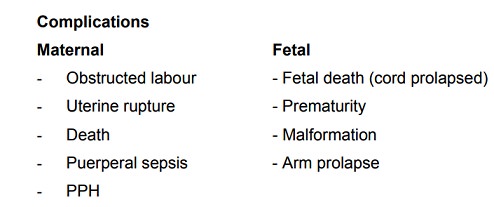
Complications
·
Obstructed labour
·
Cord prolapse
·
Facial bruising
·
Cerebral haemorrhage & Maternal trauma
5. Unstable lie
Definition:-When the lie is found to vary,
breech, vertex orshoulder, presenting from one examination to another after 36th weeks of pregnancy.
Causes
·
Lax uterine muscles
·
Multiparity
·
Poly hydraminous
Management
Admission in hospital at the 36-37 week and remain in the hospital until
delivery.
Attempts are made by the obstetrician to correct the abnormal
presentation by external version. If unsuccessful, caesarian section is
considered. Some times AROM is done after correcting the transverse lie to
ensure that the woman goes into labour with vertex presentation. An oxytocic
drip is usually given after version.
·
Extreme caution and close observation is mandatory throughout labour.
·
Monitoring of Fetal Heart Beat frequently is very important
·
The bladder and the rectum should be emptied to facilitate
·
preservation of the longitudinal lie.
6. Compound or Complex Presentation
Definition: - When a hand or occasionally of
foot, liesalong side the head. This tends to occur with a small fetus or roomy
pelvis seldom is difficulty encountered except in cases where it is associated
with a flat pelvis. On rare occasions head, hand & foot are felt in the
vagina, a serious situation which usually occurs with a dead fetus.
If diagnosed during the first stage of labour, attempt could be made to
push the arm up words over the baby’s face. If during the second stage hold the
hand back directing it over the face.
7. Occupition Posteririor Position
It is a malposition of the head, occurs in 13% of the vertex
presentations. Head is deflexed-larger diameter present.
Causes
Direct cause is unknown but associated with
·
Pendulous abdomen
·
Abnormal pelvis, Androld, Anthropoid, flat sacrum
·
The placenta is in anterior wall
Diagnose
Inspection
Deep hollow between head and lower limbs
Palpation
The fetal head is found on one side
The limbs are infornt and give hollowing above the head. There is a
saucer like depression around the umbilicus. There is a bulge like full bladder
occiput and sinciput are at the same level. Limbs are found on both sides.
Auscultation
Fetal Heart is heard in the flanks and descends down
Vaginal examination
·
Membranes may rupture early
·
If infant may protrude through cervix as a finger like fore water or
fill up the upper vagina
·
Due to deflection, anterior fontanel is felt in the anterior part of the
pelvis near ileo pectineal eminence
Out Come
·
If the flexion of the head increases the occiput strikes the pelvic
floor and rotates anteriorly (ROP) to 45 then to 900 rotation and dilvered normally.
·
If the flexion remains incomplete, the rotation of the head takes place
posteriorly brings the occiput in the hollow of the sacrum. This is known as
short rotation.In this case the baby is born by face to Pubis.
·
Some times the
long rotation of
occipitoposterior is arrested and
the head is left in the Occipito- lateral position in the cavity of the pelvis.
Occipito frontal diameter is caught at the narrow spinous diameter of
the outlet.This is known as deep transverse arrest or persistent
occiptoposterior. The delivery could be by rotation of the head to anterior or
by cesarean section.
Management
Encourage the mother to lie on the side where the fetus lies. Patient
may have sever back pain analgesics may be given. Retention of urine is common
catheterization is necessary. Patient feels the need to bear down before fully
dilation. Two-third of cases will deliver normally.12% will deliver face to
pubis. If the ischial spines are prominent the internal rotation may
interrupted caesarian section is recommended.
Identifying the ear by the root of the pinna (right or left) manual
rotation can be done by, keep the right hand on the head and left on the
abdomen and rotate than forceps delivery is performed.
Related Topics