Immunology - Lymphoid organs | 12th Zoology : Chapter 8 : Immunology
Chapter: 12th Zoology : Chapter 8 : Immunology
Lymphoid organs
Lymphoid
organs
Immune system of an
organism consists of several structurally and functionally different organs and
tissues that are widely dispersed in the body. The organs involved in the
origin, maturation and proliferation of lymphocytes are called lymphoid
organs (Fig. 8.3). Based on
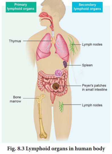
their functions, they
are classified into primary or central lymphoid organs and
secondary or peripheral lymphoid organs. The primary lymphoid organs
provide appropriate environment for lymphocytic maturation. The secondary lymphoid
organs trap antigens and make it available for mature lymphocytes, which can
effectively fight against these antigens.
Primary lymphoid organs
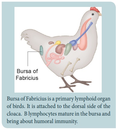
![]() Bursa of
Fabricius of birds, bone marrow and
thymus gland of mammals constitute the primary lymphoid organs involved in the
production and early selection of lymphocytes. These lymphocytes become
dedicated to a particular antigenic specificity. Only when the
lymphocytes mature in the primary lymphoidal organs, they become immunocompetent
cells. In mammals, B cell maturation occurs in the bone marrow and T
cells maturation occurs in the thymus.
Bursa of
Fabricius of birds, bone marrow and
thymus gland of mammals constitute the primary lymphoid organs involved in the
production and early selection of lymphocytes. These lymphocytes become
dedicated to a particular antigenic specificity. Only when the
lymphocytes mature in the primary lymphoidal organs, they become immunocompetent
cells. In mammals, B cell maturation occurs in the bone marrow and T
cells maturation occurs in the thymus.
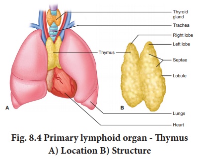
Thymus
The thymus is a flat and
bilobed organ located behind the sternun, above the heart. Each lobe of the
thymus contains numerous lobules, separated from each other by connective
tissue called septa. Each lobule is differentiated into two compartments, the
outer compartment or outer cortex, is densely packed with immature T
cells called thymocytes, whereas the inner compartment or medulla is sparsely
populated with thymocytes. One of its main secretions is the hormone thymosin.
It stimulates the T cell to become mature and immunocompetent. By the
early teens, the thymus begins to atrophy and is replaced by adipose tissue (Fig.
8.4). Thus thymus is most active during the neonatal and
pre-adolescent periods.
Bone marrow
Bone marrow is a
lymphoid tissue found within the spongy portion of the bone. Bone marrow
contains stem cells known as haematopoietic cells. These cells have the
potential to multiply through cell division and either remain as stem cells or
differentiate and mature into different kinds of blood cells.
Secondary or peripheral lymphoid organs
In secondary or
peripheral lymphoid organs, antigen is localized so that it can be effectively
exposed to mature lymphocytes. The best examples are lymph nodes, appendix,
Peyer’s patches of gastrointestinal tract, tonsils, adenoids, spleen, MALT
(Mucosal-Associated Lymphoid Tissue), GALT (Gut-Associated Lymphoid
Tissue), BALT(Bronchial/Tracheal-Associated Lymphoid Tissue).
Peyer’s patches are oval-shaped areas
of thickened tissue that are embedded in the mucus-secreting lining of the
small intestine of humans and other vertebrate animals. Peyer’s patches contain
a variety of immune cells, including macrophages, dendritic cells, T cells, and
B cells.
The tonsils (palatine tonsils) are a pair
of soft tissue masses located at the back of the throat (pharynx). The tonsils
are part of the lymphatic system, which help to fight infections. They stop
invading germs including bacteria and viruses.
Spleen is a secondary lymphoid organ located in the upper part of the abdominal cavity close to
the diaphragm. Spleen contains B and T cells. It brings humoral and cell
mediated immunity.
Lymph node
Lymph node is a small
bean-shaped structure and is part of the body’s immune system. It is the first
one to encounter the antigen that enters the tissue spaces. Lymph nodes
filter and trap substances that travel through the lymphatic fluid. They are
packed tightly with white blood cells, namely lymphocytes and macrophages.
There are hundreds of lymph nodes found throughout the body. They are connected
to one another by lymph vessels. Lymph is a clear, transparent,
colourless, mobile and extracellular fluid connective tissue. As the lymph
percolates through the lymph node, the particulate antigen brought in by the
lymph will be trapped by the phagocytic cells, follicular and
interdigitating dendritic cells.
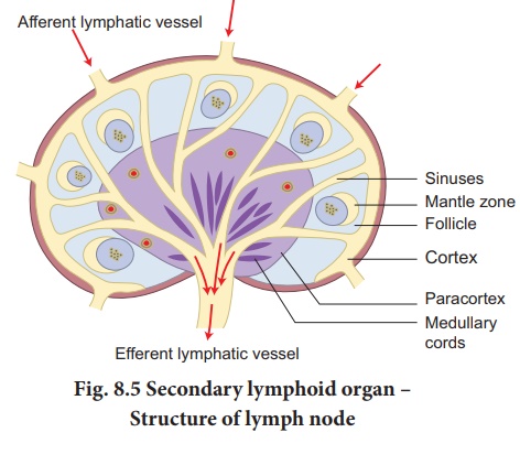
Lymph node has three
zones (Fig. 8.5). They are the cortex, paracortex and medulla.
The outer most layer of the lymph node is called cortex, which consists of
B-lymphocytes, macrophages, and follicular dendritic cells. The paracortex
zone is beneath the cortex, which is richly populated by T lymphocytes and
interdigitating dendritic cells. The inner most zone is called the medulla which
is sparsely populated by lymphocytes, but many of them are plasma cells, which
actively secrete antibody molecules. As the lymph enters, it slowly percolates
through the cortex, paracortex and medulla, giving sufficient chance for the
phagocytic cells and dendritic cells to trap the antigen brought by the lymph.
The lymph leaving a node carries enriched antibodies secreted by the medullary
plasma cells against the antigens that enter the lymph node.
Sometimes visible swelling of lymph nodes occurs due to active immune response
and increased concentration of lymphocytes. Thus swollen lymph nodes may signal
an infection. There are several groups of lymph nodes. The most frequently
enlarged lymph nodes are found in the neck, under the chin, in the armpits and
in the groin.
The mucosa-associated lymphoid tissue (MALT) is a diffuse system of small concentrations of lymphoid
tissue in the alimentary, respiratory and urino-genital tracts. MALT is populated by lymphocytes such
as T and B cells, as well as plasma cells and macrophages, each of which is
well situated to encounter antigens passing through the mucosal epithelium.
Gut-associated lymphoid tissue (GALT) is a component of the mucosa-associated lymphoid tissue (MALT) which works in the immune system
to protect the body from invasion in the gut.
Bronchus Associated Lymphoid Tissues (BALT) also a component of MALT is made of lymphoid tissue
(tonsils, lymph nodes, lymph follicles) is found in the respiratory mucosae
from the nasal cavities to the lungs.
![]()
Cells of the immune system
The immune system is
composed of many interdependent cells that protect the body from microbial
infections and the growth of tumour cells. The cellular composition of adult
human blood is given in Table 8.4.
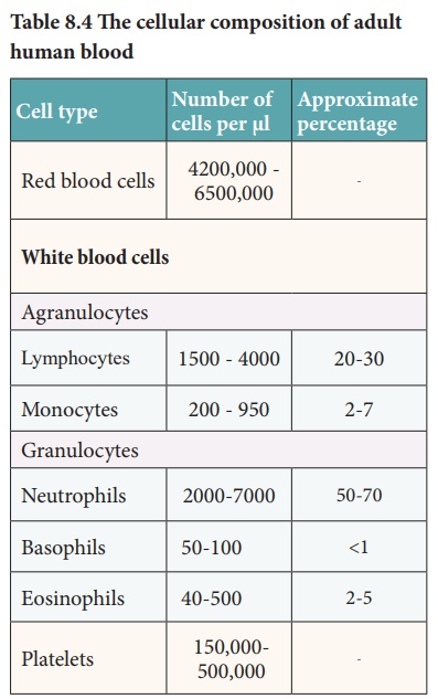
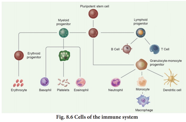
All these cells are
derived from pluripotent haematopoetic stem cells. Each stem cell has the capacity
to produce RBC, WBC and platelets.
The only cells capable
of specifically recognising and producing an immune response are the
lymphocytes. The other types of white blood cells play an important role in non
specific immune response, antigen presentation and cytokine production.
Lymphocytes
About 20-30% of the
white blood cells are lymphocytes. They have a large nucleus filling most of
the cell, surrounded by a little cytoplasm. The two main types of lymphocytes
are B and T lymphocytes. Both these are produced in the bone marrow. B
lymphocytes (B cells) stay in the bone marrow until they are mature. Then they
circulate around the body. Some remain in the blood, while others accumulate in
the lymph nodes and spleen. T lymphocytes leave the bone marrow and mature in
the thymus gland. Once mature, T cells also accumulate in the same areas of the
body as B cells. Lymphocytes have receptor proteins on their surface. When
receptors on a B cell bind with an antigen, the B cell becomes activated and
divides rapidly to produce plasma cells. The plasma cells produce antibodies.
Some B cells do not produce antibodies but become memory cells. These cells are
responsible for secondary immune response. T lymphocytes do not produce
antibodies. They recognize antigen-presenting cells and destroy them. The two
important types of T cells are Helper T cells and Killer T cells. Helper T
cells release a chemical called cytokine which activates B cells. Killer cells
move around the body and destroy cells which are damaged or infected (Fig.
8.6). ![]()
![]()
Apart from these cells
neutrophils and monocytes destroy foreign cells by phagocytosis. Monocytes when
they mature into large cells, they are called macrophages which perform
phagocytosis on any foreign organism.
Dendritic cells are
called so because its covered with long, thin membrane extensions that resemble
dendrites of nerve cells. These cells present the antigen to T-helper cells.
Four types of dendritic cells are known. They are langerhans, interstitial
cells, myeloid and lymphoid cells
Related Topics