Chapter: Essentials of Anatomy and Physiology: The Urinary System
Kidneys - Anatomy and Physiology
KIDNEYS
The two kidneys are located in the upper abdominal cavity on either side of the vertebral column, behind the peritoneum (retroperitoneal). The upper por-tions of the kidneys rest on the lower surface of the diaphragm and are enclosed and protected by the lower rib cage (see Fig. 18–1). The kidneys are embedded in adipose tissue that acts as a cushion and is in turn covered by a fibrous connective tissue mem-brane called the renal fascia, which helps hold the kidneys in place.
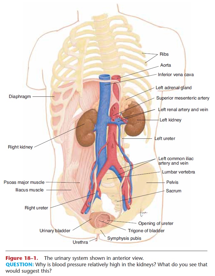
Each kidney has an indentation called the hilus on its medial side. At the hilus, the renal artery enters the kidney, and the renal vein and ureter emerge. The renal artery is a branch of the abdominal aorta, and the renal vein returns blood to the inferior vena cava (see Fig. 18–1). The ureter carries urine from the kidney to the urinary bladder.
INTERNAL STRUCTURE OF THE KIDNEY
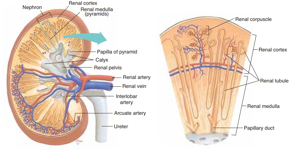
In a coronal or frontal section of the kidney, three areas can be distinguished (Fig. 18–2). The lateral and middle areas are tissue layers, and the medial area at the hilus is a cavity. The outer tissue layer is called the renal cortex; it is made of renal corpuscles and convo-luted tubules. These are parts of the nephron and are described in the next section. The inner tissue layer is the renal medulla, which is made of loops of Henle and collecting tubules (also parts of the nephron). The renal medulla consists of wedge-shaped pieces called renal pyramids. The tip of each pyramid is its apex or papilla.
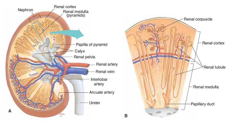
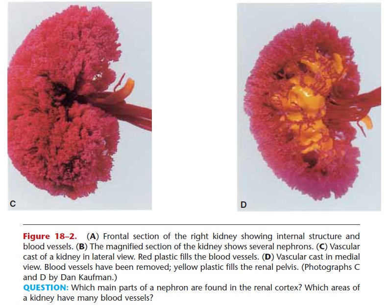
Figure 18–2. (A) Frontal section of the right kidney showing internal structure and blood vessels. (B) The magnified section of the kidney shows several nephrons. (C) Vascular cast of a kidney in lateral view. Red plastic fills the blood vessels. (D) Vascular cast in medial view. Blood vessels have been removed; yellow plastic fills the renal pelvis. (Photographs C and D by Dan Kaufman.)
QUESTION: Which main parts of a nephron are found in the renal cortex? Which areas of a kidney have many blood vessels?
The third area is the renal pelvis; this is not a layer of tissues, but rather a cavity formed by the expansion of the ureter within the kidney at the hilus. Funnel-shaped extensions of the renal pelvis, called calyces (singular: calyx), enclose the papillae of the renal pyr-amids. Urine flows from the renal pyramids into the calyces, then to the renal pelvis and out into the ureter.
THE NEPHRON
The nephron is the structural and functional unit of the kidney. Each kidney contains approximately 1 mil-lion nephrons. It is in the nephrons, with their associ-ated blood vessels, that urine is formed. Each nephron has two major portions: a renal corpuscle and a renal tubule. Each of these major parts has further subdivi-sions, which are shown with their blood vessels in Fig. 18–3.
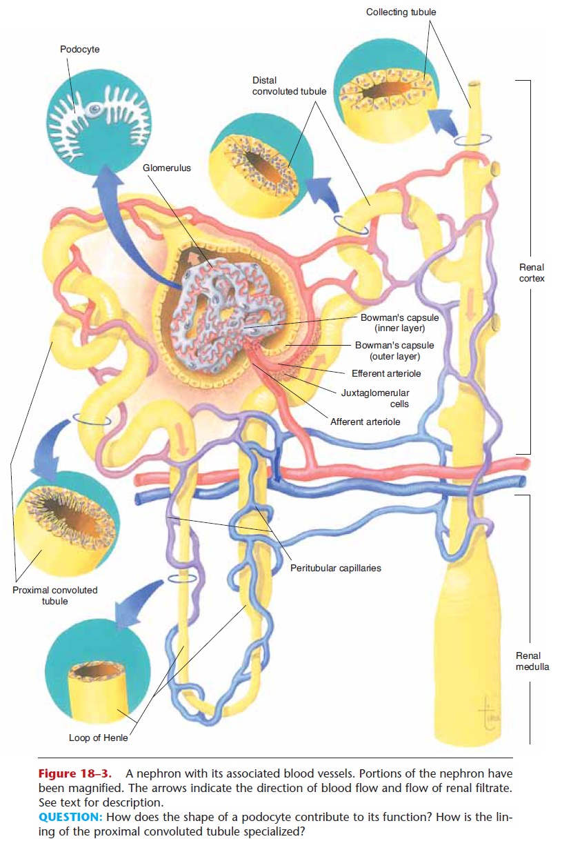
Figure 18–3. A nephron with its associated blood vessels. Portions of the nephron have been magnified. The arrows indicate the direction of blood flow and flow of renal filtrate. See text for description.
QUESTION: How does the shape of a podocyte contribute to its function? How is the lin-ing of the proximal convoluted tubule specialized?
Renal Corpuscle
A renal corpuscle consists of a glomerulus surroun-ded by a Bowman’s capsule. The glomerulus is a cap-illary network that arises from an afferent arteriole and empties into an efferent arteriole. The diameter of the efferent arteriole is smaller than that of the afferent arteriole, which helps maintain a fairly high blood pressure in the glomerulus.
Bowman’s capsule (or glomerular capsule) is the expanded end of a renal tubule; it encloses the glomerulus. The inner layer of Bowman’s capsule is made of podocytes; the name means “foot cells,” and the “feet” of the podocytes are on the surface of the glomerular capillaries. The arrangement of podocytes creates pores, spaces between adjacent “feet,” which make this layer very permeable. The outer layer of Bowman’s capsule has no pores and is not permeable. The space between the inner and outer layers of Bowman’s capsule contains renal filtrate, the fluid that is formed from the blood in the glomerulus and will eventually become urine.
Renal Tubule
The renal tubule continues from Bowman’s capsule and consists of the following parts: proximal convo-luted tubule (in the renal cortex), loop of Henle (or loop of the nephron, in the renal medulla), and distal convoluted tubule (in the renal cortex). The distalconvoluted tubules from several nephrons empty into a collecting tubule. Several collecting tubules then unite to form a papillary duct that empties urine into a calyx of the renal pelvis.
Cross-sections of the parts of the renal tubule are shown in Fig. 18–3. Notice how thin the walls of the tubule are, and also the microvilli in the proximal con-voluted tubule. These anatomic characteristics provide for efficient exchanges of materials, as you will see.
All parts of the renal tubule are surrounded by peritubular capillaries, which arise from the efferent arteriole. The peritubular capillaries will receive the materials reabsorbed by the renal tubules; this is described in the section on urine formation.
BLOOD VESSELS OF THE KIDNEY
The pathway of blood flow through the kidney is an essential part of the process of urine formation. Blood from the abdominal aorta enters the renal artery, which branches extensively within the kidney into smaller arteries (see Fig. 18–2). The smallest arteries give rise to afferent arterioles in the renal cortex (see Fig. 18–3). From the afferent arterioles, blood flows into the glomeruli (capillaries), to efferent arterioles, to peritubular capillaries, to veins within the kidney, to the renal vein, and finally to the inferior vena cava Notice that in this pathway there are two sets of cap-illaries, and recall that it is in capillaries that exchanges take place between the blood and surrounding tissues. Therefore, in the kidneys there are two sites of ex-change. The exchanges that take place between the nephrons and the capillaries of the kidneys will form urine from blood plasma.
Figure 18–2 shows two views of a vascular cast of a kidney; the shape of the blood vessels has been pre-served in red plastic. You can see how dense the vas-culature of a kidney is, and most of these vessels are capillaries.
Related Topics