Chapter: Ophthalmology: Retina
Hypertensive Retinopathy and Sclerotic Changes
Hypertensive Retinopathy and Sclerotic Changes
Definition
Arterial changes in hypertension are primarily
caused by vasospasm; in arterio-sclerosis they are the result of thickening of
the wall of the arteriole.
Epidemiology:
Arterial hypertension in particular figures prominently
inclinical settings.
Vascular changes due to arterial hypertension
are the most frequent cause of retinal vein occlusion.
Pathogenesis:
High blood pressure can cause breakdown of the
blood-retinabarrier or obliteration of capillaries. This results in
intraretinal bleeding, cot-ton-wool spots, retinal edema, or swelling of the
optic disk.
Symptoms:
Patients with high blood pressure frequently suffer from
head-ache or eye pain. Impaired vision or loss of visual acuity only occurs in
stage III or IV hypertensive vascular changes. Arteriosclerosis does not
exhibit any ocular symptoms.
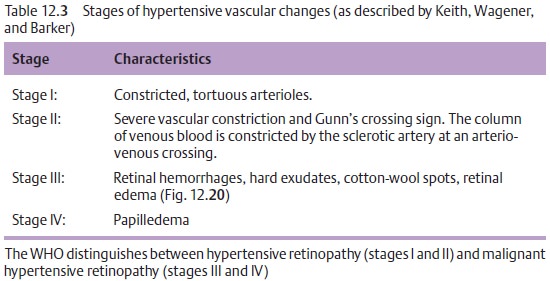
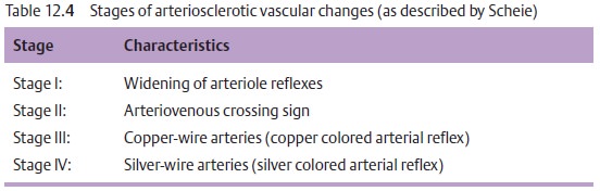
Diagnostic considerations:
Hypertensive and arteriosclerotic changes in thefundus are
diagnosed by ophthalmoscopy, preferably with the pupil dilated (Tables 12.3 and 12.4). Changes in the retinal vasculature are frequent find-ings;
choroidal infarctions are rare in acute hypertension (Elschnig’s spots:
circumscribed atrophy and proliferation of pigment epithelium in the infarcted
area).
Differential diagnosis:
Ophthalmoscopy should be performed to excludeother vascular
retinal disorders such as diabetic retinopathy. Diabetic reti-nopathy is
primarily characterized by parenchymal and vascular changes; a differential
diagnosis is made by confirming or excluding the systemic under-lying disorder.
Treatment:
Treating the underlying disorder is crucial where fundus changesdue to arterial retinopathy are present. Blood pressure should be reduced to below 140/90 mm Hg. Fundus changes due to arteriosclerosis are untreat-able.
Prophylaxis:
Regular blood pressure monitoring and ophthalmoscopicexamination
of the fundus are required to minimize the risk of complications (see below).
Clinical course and complications:
Sequelae of arteriosclerotic and hyper-tensive vascular changes
include retinal artery and vein occlusion and the for-mation of macroaneurysms
that can lead to vitreous hemorrhage. In the pres-ence of papilledema, the
subsequent atrophy of the optic nerve can produce lasting and occasionally
severe loss of visual acuity.
Prognosis:
In some cases, the complications described above are unavoidable
despite well controlled blood pressure.
Related Topics