Chapter: 11th Zoology : Chapter 6 : Respiration
Human Respiratory System - Respiratory organs in various organisms
Respiratory organs in various organisms.
Different
animals have different organs for exchange of gases, depending upon their
habitats and levels of organization. The amount of dissolved oxygen is very low
in water compared to the amount of oxygen in the air. So the rate of breathing
in aquatic organisms is much faster than land animals.
In
animals like sponges, coelenterates and flatworms exchange of gases takes place
through the body surface by simple diffusion. Earthworms use their moist skin,
whereas insects have tracheal tubes. Gills are used as respiratory organs in
most of the aquatic Arthropods and Molluscs. Among verterbrates, fishes use gills
whereas amphibians, reptiles, birds and mammals have well vascularised lungs.
Frogs spend most of their time in water and also use their moist skin for
respiration along with lungs.
Human Respiratory System
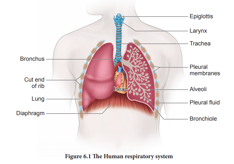
The
respiratory system includes the external nostrils, nasal cavity, the pharynx,
the larynx, the trachea, the bronchi and bronchioles and the lungs which
contain the alveoli (Figure 6.1). The parts starting from the external nostrils
up to the terminal bronchioles constitute the conducting zone, whereas the
alveoli and the ducts are called the respiratory zone. The parts of the
conducting zone, humidifies and warms the incoming air.
In human beings, air enters the upper respiratory tract through the external nostrils. The air passing through the nostrils is filtered by fine hairs and mucus lining the passage. The external nostrils lead to the nasal chamber which opens into the nasopharynx which opens through the glottis of the larynx region into the trachea. The ciliated epithelial cells lining the trachea, bronchi and bronchioles secrete mucus. Mucus membrane lining the airway contains goblet cells which secrete mucus, a slimy material rich in glycoprotein. Microorganisms and dust particles attach in the mucus films and are carried upwards to pass down the gullet during normal swallowing. During swallowing a thin elastic flap called epiglottis prevents the food from entering into the larynx and avoids choking of food.
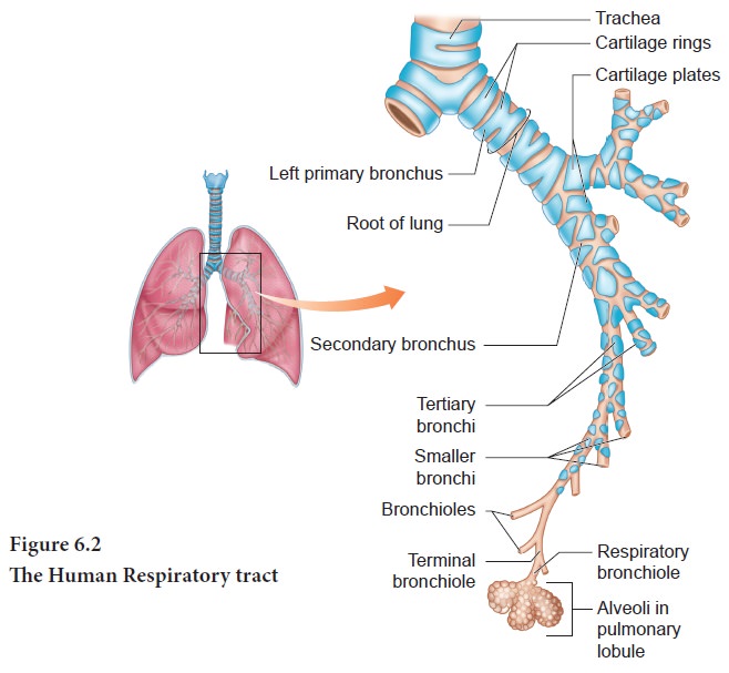
The
trachea is semiflexible tube supported by multiple cartilaginous rings which
extends up to the midthoracic cavity and at the level of the 5th thoracic
vertebra where it divides into right and left primary bronchi, one bronchus to
each lung. Within the lungs the bronchi divides repeatedly into secondary and
tertiary bronchi and further divides into terminal bronchioles and respiratory
bronchioles.
Bronchi
have ‘C’ shaped curved cartilage plates to ensure that the air passage does not
collapse or burst as the air pressure changes during breathing.
The
bronchioles- are without cartilaginous rings and have rigidity that prevent
them from collapsing but are surrounded by smooth muscle which contracts or
relaxes to -adjust the diameter of these airways.
The fine respiratory bronchioles -terminate into highly vascularised thin walled pouch like air sacs called alveoli meant for gaseous exchange (Figure 6.2, 6. 3). The diffusion membrane of alveolus is made up of three layers – the thin squamous epithelial cells of the alveoli, the endothelium of the alveolar -capillaries and the basement substance- found in between them. The thin squamous epithelial cells of the alveoli are composed of Type I and Type II cells. Type I cells are very thin so that gases can diffuse rapidly through them.
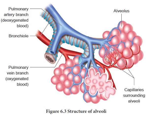
Type II
cells are thicker, synthesize and secrete- a substance called Surfactant.
The lungs
are light spongy tissues enclosed in the thoracic cavity surrounded by an
airtight space. The thoracic cavity is bound dorsally by the vertebral column
and ventrally by the sternum, laterally by the ribs and on the lower side by
the dome shaped diaphragm.
The lungs
are covered by double walled pleural membrane containing a -several layers of
elastic connective tissues and capillaries, which encloses the pleural fluid.
Pleural fluid reduces friction when the lungs expand and contract.
Characteristic features of respiratory surface:
•
surface area must be very large and richly supplied
with blood vessels
•
should be extremely thin and kept moist
•
should be in direct contact with the environment
•
should be permeable to respiratory gases
The steps
involved in respiration are
i. The
exchange of air between the atmosphere and the lungs.
ii.
The
exchange of O2
and CO2 between- the lungs and the blood.
iii.
Transport of O2 and CO2 by
the blood.
iv.
Exchange of gases between the blood and the cells.
v.
Uptake of O2 by the cells for various
activities and the release of CO2.
Related Topics