Chapter: Human Nervous System and Sensory Organs : Brain Stem and Cranial Nerves
Histochemistry of the Brain Stem
Histochemistry of the Brain Stem
Different
regions of the brain stem are characterized by different contents in chemical
substances. The delimitation of areas according to their chemical composition
is called chemoarchitectonics.
Substances can be demonstrated by quantitative chemical analysis after
homogenization of the brain tissue, or by treating histological sections with
certain chemicals that make it possible to show the exact localization of a
substance in the tissue. The methods com-plement each other.
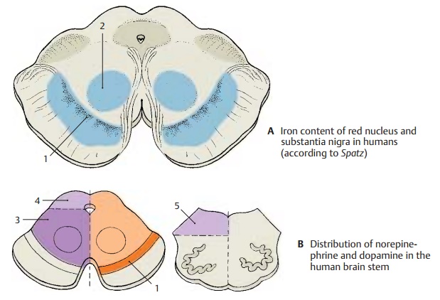
Iron was one of the first substances forwhich different
distributions were demon-strated. By means of the Berlin blue reac-tion, a high
iron content can be demon-strated in the substantia nigra (A1) and in the pallidum, while a lower iron content is found in the
red nucleus (A2), in the dentate
nucleus of the cerebellum, and in the stri-atum. The iron is contained in
neurons and glial cells in the form of small particles. This high iron content
is a characteristic of the nuclei that make up the extrapyramidal system.
Neurotransmitter substances and
theenzymes required for their synthesis and degradation show marked regional
varia-tions. While catecholaminergic
and sero-toninergic neurons form
specific nuclei inthe tegmentum, the motor nuclei of cranial nerves are
characterized by a high content in acetylcholine
and acetylcholineesterase.
Quantitative chemical analysis ofbrain tissue yields a relatively high content
of norepinephrine in the tegmentum of
the midbrain (B3), but a
considerably lower content in the tectum (B4)
and in the teg-mentum of the medulla oblongata (B5). The content of dopamine
is particularly high in the substantia nigra (B1) and very low in the rest of the brain stem.
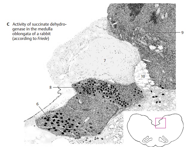
Metabolic enzymes (C) also show regionalvariations in their distribution. Activity of oxidative enzymes is generally higher in
graymatter than in white matter. In the brainstem, activity is particularly
high in the cranial nerve nuclei, the lower portion of the olive, and the
pontine nuclei. The differ-ences refer not only to the individual areas but
also to the localization of enzyme activ-ity within the cell bodies (somatic type) or in the neuropil (dendritic type).

Neuropil. The substance between the
cellbodies, which appears amorphous in Nissl-stained material, is called the neuropil.
It consists mainly of dendrites and also of axons and glial processes. The
majority of all synaptic contacts are found in the neuropil.
The
distribution in the medulla oblongata of succinate
dehydrogenase (an enzyme of thecitric acid cycle) serves as an example for
different localizations of an oxidative meta-bolic enzyme within the tissue: in
the oculo-motor nucleus (C6), its activity in the peri-karya and
in the neuropil is high, while it is low at both locations in the solitary nucleus (C7). In the posterior nucleus
of the vagusnerve (C8), the cell
bodies contrast with theneuropil owing to their high activity. By comparison,
the highly active neuropil in the gracile
nucleus (C9) lets the poorly
re-acting perikarya appear as light spots. Fiber tracts (for example, the solitary tract) (C10) show very low activity. The distribution of enzymes is
characteristic for each nuclear area and is referred to as the enzyme pattern.
Related Topics