Chapter: Medical Physiology: The Sense of Hearing
Hearing Abnormalities: Types of Deafness
Hearing Abnormalities
Types of Deafness
Deafness is usually divided into two types: (1) that caused by impairment of the cochlea or impairment of the auditory nerve, which is usually classified as “nerve deafness,” and (2) that caused by impairment of the physical structures of the ear that conduct sound itself to the cochlea, which is usually called “conduction deafness.”
If either the cochlea or the auditory nerve is destroyed, the person becomes permanently deaf. However, if the cochlea and nerve are still intact but the tympanum-ossicular system has been destroyed or ankylosed (“frozen” in place by fibrosis or calcification), sound waves can still be conducted into the cochlea by means of bone conduction from a sound generator applied to the skull over the ear.
Audiometer. To determine the nature of hearing dis-abilities, the “audiometer” is used. Simply an earphone connected to an electronic oscillator capable of emitting pure tones ranging from low frequencies to high fre-quencies, the instrument is calibrated so that zero-intensity-level sound at each frequency is the loudness that can barely be heard by the normal ear. A calibrated volume control can increase the loudness above the zero level. If the loudness must be increased to 30 deci-bels above normal before it can be heard, the person is said to have a hearing loss of 30 decibels at that partic-ular frequency.
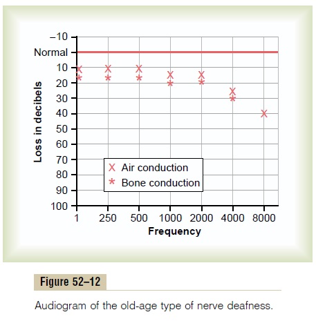
In performing a hearing test using an audiometer, one tests about 8 to 10 frequencies covering the auditory spectrum, and the hearing loss is determined for each of these frequencies. Then the so-called audiogram is plotted, as shown in Figures 52–12 and 52–13, depicting hearing loss at each of the frequencies in the auditory spectrum. The audiometer, in addition to being equipped with an earphone for testing air conduction by the ear, is equipped with a mechanical vibrator for testing bone conduction from the mastoid process of the skull into the cochlea.
Audiogram in Nerve Deafness. In nerve deafness—which includes damage to the cochlea, the auditory nerve, or the central nervous system circuits from the ear—the person has decreased or total loss of ability to hear sound as tested by both air conduction and bone conduction. An audiogram depicting partial nerve deaf-ness is shown in Figure 52–12. In this figure, the deaf-ness is mainly for high-frequency sound. Such deafness could be caused by damage to the base of the cochlea. This type of deafness occurs to some extent in almost all older people.
Other patterns of nerve deafness frequently occur as follows: (1) deafness for low-frequency sounds caused by excessive and prolonged exposure to very loud sounds (a rock band or a jet airplane engine), because low-frequency sounds are usually louder and more dam-aging to the organ of Corti, and (2) deafness for all frequencies caused by drug sensitivity of the organ of Corti—in particular, sensitivity to some antibiotics such as streptomycin, kanamycin, and chloramphenicol.
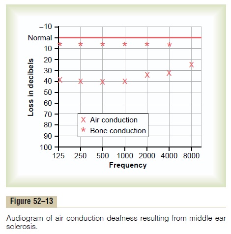
Audiogram for Middle Ear Conduction Deafness. Acommon type of deafness is caused by fibrosis in the middle ear following repeated infection or by fibrosis that occurs in the hereditary disease called otosclerosis. In either case, the sound waves cannot be transmitted easily through the ossicles from the tympanic mem-brane to the oval window. Figure 52–13 shows an audio-gram from a person with “middle ear air conduction deafness.” In this case, bone conduction is essentially normal, but conduction through the ossicular system is greatly depressed at all frequencies, but more so at low frequencies. In some instances of conduction deafness, the faceplate of the stapes becomes “ankylosed” by bone overgrowth to the edges of the oval window. In this case, the person becomes totally deaf for ossicular conduction but can regain almost normal hearing by the surgical removal of the stapes and its replacement with a minute Teflon or metal prosthesis that transmits the sound from the incus to the oval window.
Related Topics