Chapter: Medical Physiology: Secretory Functions of the Alimentary Tract
General Principles of Alimentary Tract Secretion
General Principles of Alimentary Tract Secretion
Anatomical Types of Glands
Several types of glands provide the different types of alimentary tract secretions. First, on the surface of the epithelium in most parts of the gastrointestinal tract are billions of single-cell mucous glands called simply mucous cells or sometimes gobletcells because they look like goblets. They function mainly in response to local irri-tation of the epithelium: they extrude mucus directly onto the epithelial surface to act as a lubricant that also protects the surfaces from excoriation and digestion.
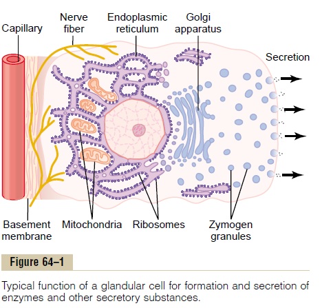
Second, many surface areas of the gastrointestinal tract are lined by pits that rep-resent invaginations of the epithelium into the submucosa. In the small intestine, these pits, calledcrypts of Lieberkühn, are deep and contain specialized secretory cells. One of these cells is shown in Figure 64–1.
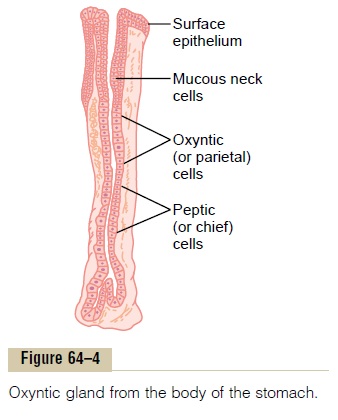
Third, in the stomach and upper duodenum are large numbers of deep tubularglands. A typical tubular gland can be seen in Figure 64–4, which shows an acid- andpepsinogen-secreting gland of the stomach (oxyntic gland).
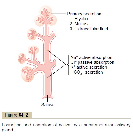
Fourth, also associated with the alimentary tract are several complex glands—the salivary glands, pancreas, and liver—that provide secretions for digestion or emul-sification of food. The liver has a highly specialized structure. The salivary glands and the pancreas are compound acinous glands of the type shown in Figure 64–2. These glands lie outside the walls of the alimentary tract and, in this, differ from all other alimentary glands. They contain millions of acini lined with secreting glandular cells; these acini feed into a system of ducts thatfinally empty into the alimentary tract itself.
Basic Mechanisms of Stimulation of the Alimentary Tract Glands
Effect of Contact of Food with the Epithelium—Function of Enteric Nervous Stimuli. Themechanical presence of food in a particular segment of the gastrointestinal tract usually causes the glands of that region and often of adjacent regions to secrete moderate to large quantities of juices. Part of this local effect, especially the secretion of mucus by mucous cells, results from direct contact stimulation of the surface glandular cells by the food.
In addition, local epithelial stimulation also acti-vates the enteric nervous system of the gut wall. The types of stimuli that do this are (1) tactile stimulation, (2) chemical irritation, and (3) distention of the gut wall. The resulting nervous reflexes stimulate both the mucous cells on the gut epithelial surface and the deep glands in the gut wall to increase their secretion.
Autonomic Stimulation of Secretion Parasympathetic Stimulation. Stimulation of theparasympathetic nerves to the alimentary tract almost invariably increases the rates of alimentary glandula secretion. This is especially true of the glands in the upper portion of the tract (innervated by the glos-sopharyngeal and vagus parasympathetic nerves) such as the salivary glands, esophageal glands, gastric glands, pancreas, and Brunner’s glands in the duode-num. It is also true of some glands in the distal portion of the large intestine, innervated by pelvic parasym-pathetic nerves. Secretion in the remainder of the small intestine and in the first two thirds of the large intestine occurs mainly in response to local neural and hormonal stimuli in each segment of the gut.
Sympathetic Stimulation. Stimulation of the sympa-thetic nerves going to the gastrointestinal tract causes a slight to moderate increase in secretion by some of the local glands. But sympathetic stimulation also results in constriction of the blood vessels that supply the glands. Therefore, sympathetic stimulation can have a dual effect: First, sympathetic stimulation alone usually slightly increases secretion. But, second, if parasympathetic or hormonal stimulation is already causing copious secretion by the glands, superimposed sympathetic stimulation usually reduces the secretion, sometimes significantly so, mainly because of vaso-constrictive reduction of the blood supply.
Regulation of GlandularSecretion by Hormones. In the stomach and intestine, several different gastrointestinalhormones help regulate the volume and character ofthe secretions. These hormones are liberated from the gastrointestinal mucosa in response to the presence of food in the lumen of the gut. The hormones then are absorbed into the blood and carried to the glands, where they stimulate secretion. This type of stimula-tion is particularly valuable to increase the output of gastric juice and pancreatic juice when food enters the stomach or duodenum.
Chemically, the gastrointestinal hormones are polypeptides or polypeptide derivatives.
Basic Mechanism of Secretion by Glandular Cells
Secretion of Organic Substances. Although all thebasic mechanisms by which glandular cells function are not known, experimental evidence points to the following principles of secretion, as shown in Figure 64–1.
1.The nutrient material needed for formation of the secretion must first diffuse or be actively transported by the blood in the capillaries into the base of the glandular cell.
2.Many mitochondria located inside the glandular cell near its base use oxidative energy to form adenosine triphosphate (ATP).
3.Energy from the ATP, along with appropriate substrates provided by the nutrients, is then used to synthesize the organic secretory substances; this synthesis occurs almost entirely in the endoplasmic reticulum and Golgi complex of theglandular cell. Ribosomes adherent to the reticulum are specifically responsible for formation of the proteins that are secreted.
4.The secretory materials are transported through the tubules of the endoplasmic reticulum, passing in about 20 minutes all the way to the vesicles of the Golgi complex.
5.In the Golgi complex, the materials are modified, added to, concentrated, and discharged into the cytoplasm in the form of secretory vesicles, which are stored in the apical ends of the secretory cells.
6.These vesicles remain stored until nervous or hormonal control signals cause the cells to extrude the vesicular contents through the cells’ surface. This probably occurs in the following way: The control signal first increases the cellmembrane permeability to calcium ions, andcalcium enters the cell. The calcium in turn causes many of the vesicles to fuse with the apical cell membrane. Then the apical cell membrane breaks open, thus emptying the vesicles to the exterior; this process is called exocytosis.
Water and Electrolyte Secretion. A second necessity forglandular secretion is secretion of sufficient water and electrolytes to go along with the organic substances. The following is a postulated method by which nervous stimulation causes water and salts to pass through the glandular cells in great profusion, washing the organic substances through the secretory border of the cells at the same time:
1.Nerve stimulation has a specific effect on the basal portion of the cell membrane to cause activetransport of chloride ions to the cell interior.
2.The resulting increase in electronegativity induced inside the cell by excess negatively charged chloride ions then causes positive ions such as sodium ions also to move through the cellmembrane to the interior of the cell.
3.Now, the new excess of both negative and positive ions inside the cell creates an osmotic force that causes osmosis of water to the interior, thereby increasing cell volume and hydrostatic pressure inside the cell, causing the cell itself to swell.
4.The pressure in the cell then initiates minute openings of the secretory border of the cell, causing flushing of water, electrolytes, and organic materials out of the secretory end of the glandular cell.
In support of these secretory processes have been the following findings: First, the nerve endings on glan-dular cells are principally on the bases of the cells. Second, microelectrode studies show that the normal electrical potential across the membrane at the base of the cell is between 30 and 40 millivolts, with negativ-ity on the interior and positivity on the exterior. Parasympathetic stimulation increases this polariza-tion voltage to values some 10 and 20 millivolts more negative than normal. This increase in polarization voltage lasts for 1 second or longer after the nerve signal has arrived, indicating that it is caused by move-ment of negative ions (presumably chloride ions) through the membrane to the interior of the cell, thus leading to secretion.
Although this mechanism for secretion is still partly theoretical, it does explain how it would be possible for nerve impulses to regulate secretion. Hormones acting on the cell membrane are believed also to cause similar secretory results to those caused by nervous stimulation.
Lubricating and Protective Properties of Mucus, and Importance of Mucus in the Gastrointestinal Tract
Mucus is a thick secretion composed mainly of water, electrolytes, and a mixture of several glycoproteins, which themselves are composed of large polysaccha-rides bound with much smaller quantities of protein. Mucus is slightly different in different parts of the gas-trointestinal tract, but everywhere it has several im-portant characteristics that make it both an excellent lubricant and a protectant for the wall of the gut. First, mucus has adherent qualities that make it adhere tightly to the food or other particles and to spread as a thin film over the surfaces. Second, it has sufficient body that it coats the wall of the gut and prevents actual contact of most food particles with the mucosa. Third, mucus has a low resistance for slippage, so that the particles can slide along the epithelium with great ease. Fourth, mucus causes fecal particles to adhere to one another to form the feces that are expelled during a bowel move-ment. Fifth, mucus is strongly resistant to digestion by the gastrointestinal enzymes. And sixth, the glycopro-teins of mucus have amphoteric properties, which means that they are capable of buffering small amounts of either acids or alkalies; also, mucus often contains moderate quantities of bicarbonate ions which specifi-cally neutralize acids.
In summary, mucus has the ability to allow easy slip-page of food along the gastrointestinal tract and to prevent excoriative or chemical damage to the epithe-lium. A person becomes acutely aware of the lubricat-ing qualities of mucus when the salivary glands fail to secrete saliva, because then it is difficult to swallow solid food even when it is eaten along with large amounts of water.
Related Topics