Chapter: Medical Physiology: The Autonomic Nervous System and the Adrenal Medulla
General Organization of the Autonomic Nervous System
General Organization of the Autonomic Nervous System
The autonomic nervous system is activated mainly by centers located in the spinal cord, brain stem, and hypothalamus. Also, portions of the cerebral cortex,especially of the limbic cortex, can transmit signals to the lower centers and in this way influence autonomic control.
The autonomic nervous system also often operates by means of visceralreflexes. That is, subconscious sensory signals from a visceral organ can enterthe autonomic ganglia, the brain stem, or the hypothalamus and then return subconscious reflex responses directly back to the visceral organ to control itsactivities.
The efferent autonomic signals are transmitted to the various organs of the body through two major subdivisions called the sympathetic nervous system and the parasympathetic nervous system, the characteristics and functions of which follow.
Physiologic Anatomy of the Sympathetic Nervous System
Figure 60–1 shows the general organization of the peripheral portions of the sym-pathetic nervous system. Shown specifically in the figure are (1) one of the two paravertebral sympathetic chains of ganglia that are interconnected with the spinalnerves on the side of the vertebral column, (2) twoprevertebral ganglia (the celiac and hypogastric), and (3) nerves extending from the ganglia to the different inter-nal organs.
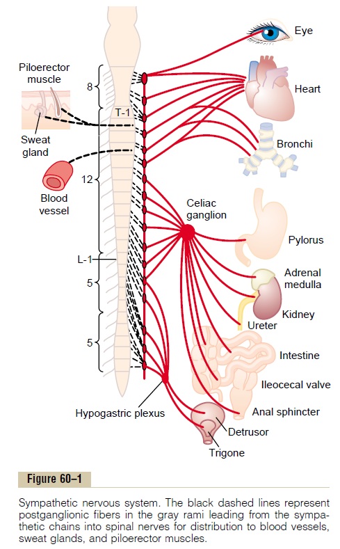
The sympathetic nerve fibers originate in the spinal cord along with spinal nerves between cord segments T-1 and L-2 and pass first into thesympathetic chain and then to the tissues and organs that are stimulated by the sympathetic nerves.
Preganglionic and Postganglionic Sympathetic Neurons
The sympathetic nerves are different from skeletal motor nerves in the following way: Each sympathetic pathway from the cord to the stimulated tissue is composed
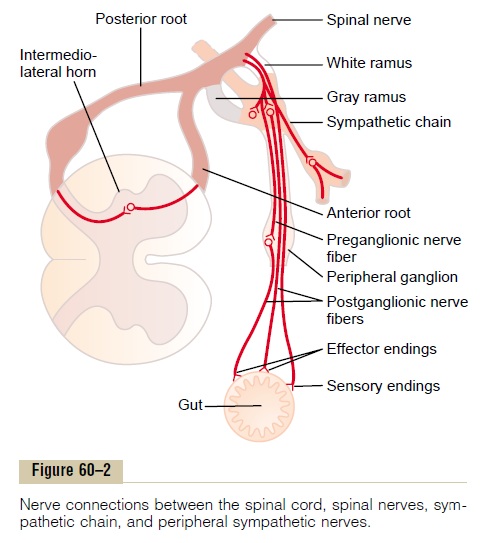
of two neurons, a preganglionic neuron and a postgan-glionic neuron, in contrast to only a single neuron in theskeletal motor pathway. The cell body of each pregan-glionic neuron lies in the intermediolateral horn of the spinal cord; its fiber passes, as shown in Figure 60–2, through an anterior rootof the cord into the corre-sponding spinal nerve.
Immediately after the spinal nerve leaves the spinal canal, the preganglionic sympathetic fibers leave the spinal nerve and pass through a white ramusinto one of the ganglia of the sympathetic chain. Then the course of the fibers can be one of the following three: (1) It can synapse with postganglionic sympathetic neurons in the ganglion that it enters; (2) it can pass upward or downward in the chain and synapse in one of the other ganglia of the chain; or (3) it can pass for variable distances through the chain and then through one of the sympathetic nerves radiating outward from the chain, finally synapsing in a peripheral sympatheticganglion.
The postganglionic sympathetic neuron thus origi-nates either in one of the sympathetic chain ganglia or in one of the peripheral sympathetic ganglia. From either of these two sources, the postganglionic fibers then travel to their destinations in the various organs.
Sympathetic Nerve Fibers in the Skeletal Nerves. Some of thepostganglionic fibers pass back from the sympathetic chain into the spinal nerves through gray rami at all levels of the cord, as shown in Figure 60–2. These sym-pathetic fibers are all very small type C fibers, and they extend to all parts of the body by way of the skeletal nerves. They control the blood vessels, sweat glands, and piloerector muscles of the hairs. About 8 per cent of the fibers in the average skeletal nerve are sympathetic fibers, a fact that indicates their great importance.
Segmental Distribution of the Sympathetic Nerve Fibers. Thesympathetic pathways that originate in the different segments of the spinal cord are not necessarily distrib-uted to the same part of the body as the somatic spinal nerve fibers from the same segments. Instead, the sym-pathetic fibers from cord segment T-1 generally pass up the sympathetic chain to terminate in the head; from T-2 to terminate in the neck; from T-3, T-4, T-5, and T-6 into the thorax; from T-7, T-8, T-9, T-10, and T-11 into the abdomen; and from T-12, L-1, and L-2 into the legs.
This distribution is only approximate and overlaps greatly.
The distribution of sympathetic nerves to each organ is determined partly by the locus in the embryo from which the organ originated. For instance, the heart receives many sympathetic nerve fibers from the neck portion of the sympathetic chain because the heart orig-inated in the neck of the embryo before translocating into the thorax. Likewise, the abdominal organs receive most of their sympathetic innervation from the lower thoracic spinal cord segments because most of the prim-itive gut originated in this area.
Special Nature of the Sympathetic Nerve Endings in the Adrenal Medullae. Preganglionic sympathetic nerve fibers pass,without synapsing, all the way from the intermedio-lateral horn cells of the spinal cord, through the sympathetic chains, then through the splanchnic nerves, and finally into the two adrenal medullae. There they end directly on modified neuronal cells that secrete epi-nephrine and norepinephrine into the blood stream.These secretory cells embryologically are derived from nervous tissue and are actually themselves postgan-glionic neurons; indeed, they even have rudimentary nerve fibers, and it is the endings of these fibers that secrete the adrenal hormones epinephrine and norepinephrine.
Physiologic Anatomy of the Parasympathetic Nervous System
The parasympathetic nervous system is shown in Figure 60–3, demonstrating that parasympathetic fibers leave the central nervous system through cranial nerves III, VII, IX, and X; additional parasympathetic fibers leave the lowermost part of the spinal cord through the second and third sacral spinal nerves and occasionally the first and fourth sacral nerves. About 75 per cent of all parasympathetic nerve fibers are in the vagus nerves (cranial nerve X), passing to the entire thoracic and abdominal regions of the body. Therefore, a physiologist speaking of the parasympathetic nervous system often thinks mainly of the two vagus nerves. The vagus nerves supply parasympathetic nerves to the heart, lungs, esophagus, stomach, entire small intestine, proximal half of the colon, liver, gallbladder, pancreas, kidneys, and upper portions of the ureters.
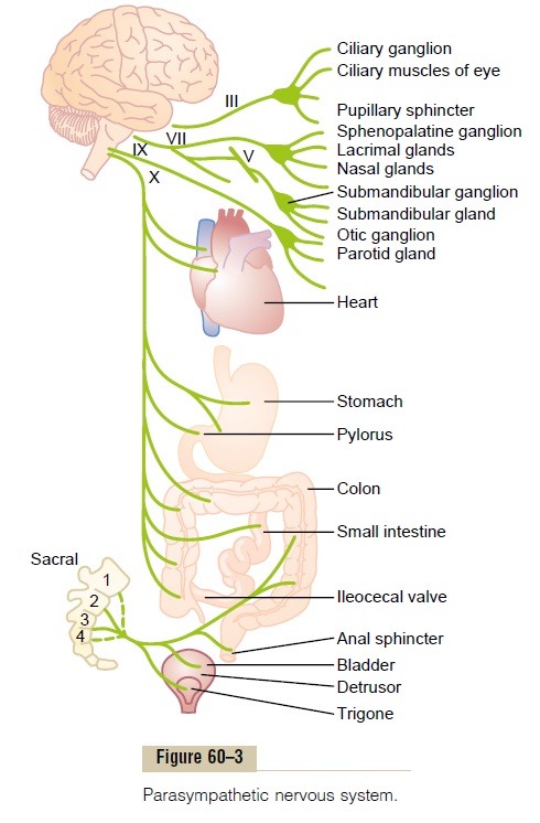
Parasympathetic fibers in the third cranial nerve go to the pupillary sphincter and ciliary muscle of the eye. Fibers from the seventh cranial nerve pass to the lacrimal, nasal, and submandibular glands. And fibers from the ninth cranial nerve go to the parotid gland.
The sacral parasympathetic fibers are in the pelvicnerves, which pass through the spinal nerve sacralplexus on each side of the cord at the S-2 and S-3 levels. These fibers then distribute to the descending colon, rectum, urinary bladder, and lower portions of the ureters. Also, this sacral group of parasympathetics sup-plies nerve signals to the external genitalia to cause erection.
Preganglionic and Postganglionic Parasympathetic Neurons.
The parasympathetic system, like the sympathetic, has both preganglionic and postganglionic neurons. However, except in the case of a few cranial parasym-pathetic nerves, the preganglionic fibers pass uninter-rupted all the way to the organ that is to be controlled. In the wall of the organ are located the postganglionicneurons. The preganglionic fibers synapse with these,and very, very short postganglionic fibers, a fraction of a millimeter to several centimeters in length, leave the neurons to innervate the tissues of the organ. This loca-tion of the parasympathetic postganglionic neurons in the visceral organ itself is quite different from the arrangement of the sympathetic ganglia, because the cell bodies of the sympathetic postganglionic neurons are almost always located in the ganglia of the sympa-thetic chain or in various other discrete ganglia in the abdomen, rather than in the excited organ itself.
Related Topics