Chapter: Essentials of Anatomy and Physiology: Blood Vessels and Circulation
General Features of Blood Vessel Structure
GENERAL FEATURES OF BLOOD VESSEL STRUCTURE
The three main types of blood vessels are arteries, capillaries, and veins. Arteries (ar′ ter-ēz) carry blood away from the heart; usually, the blood is oxygen-rich. Blood is pumped from the ventricles of the heart into large, elastic arteries, which branch repeatedly to form progressively smaller arteries. As they become smaller, the artery walls undergo a gradual transition from having more elastic tissue than smooth muscle to having more smooth muscle than elastic tissue (figure 13.1a–c). The arteries are normally classified as elastic arteries, muscular arteries, or arterioles, although they form a continuum from the largest to the smallest branches.
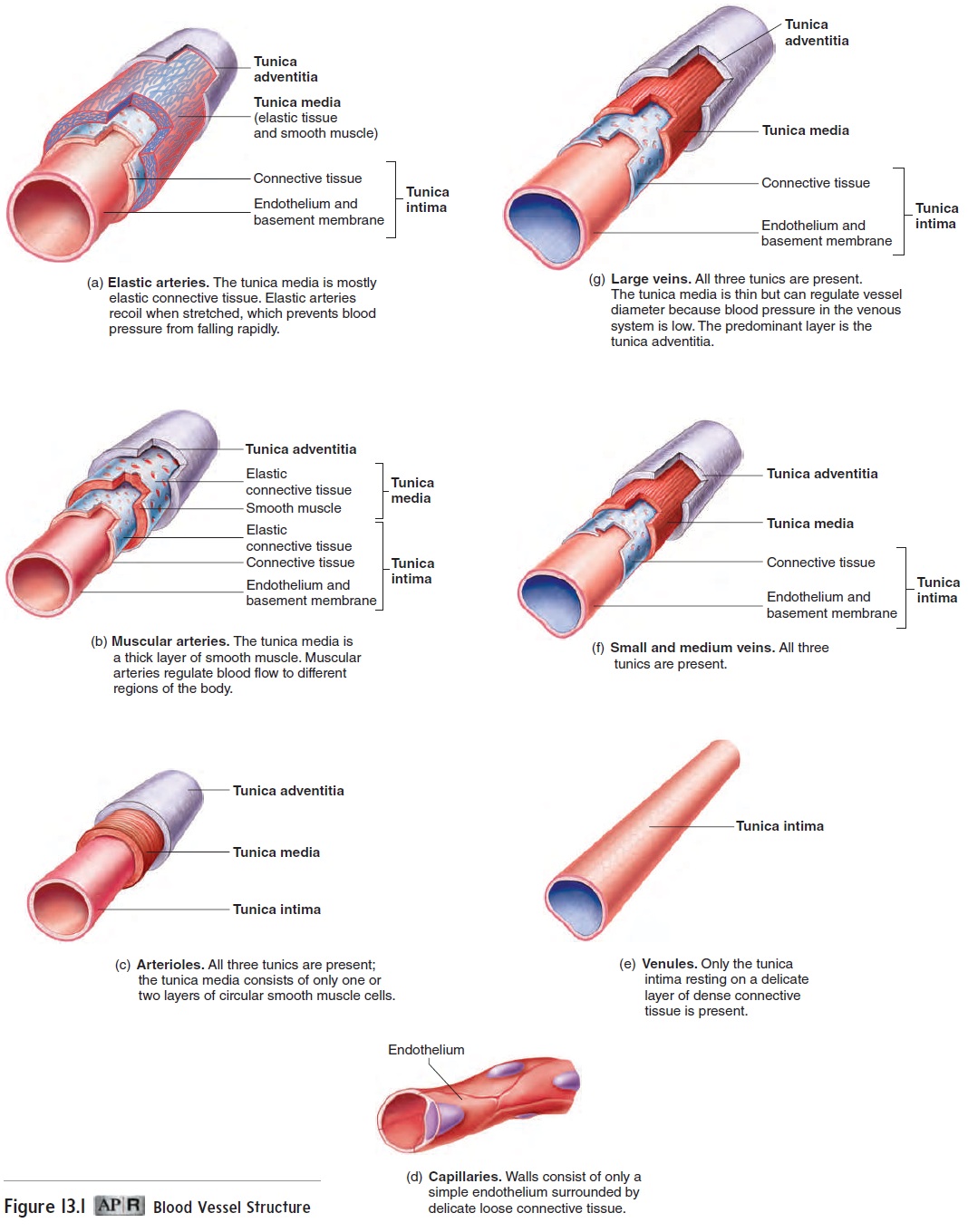
Blood flows from arterioles into capillaries (kap′ i-lār-ēz), where exchange occurs between the blood and the tissue fluid. Capillaries have thinner walls than do arteries (figure 13.1d). Blood flows through them more slowly, and there are far more of them than of any other blood vessel type.
From the capillaries, blood flows into veins. Veins (vānz) carry blood toward the heart; usually, the blood is oxygen-poor. Compared to arteries, the walls of veins are thinner and contain less elastic tissue and fewer smooth muscle cells (figure 13.1e–g). Starting at capillaries and proceeding toward the heart, small-diameter veins come together to form larger-diameter veins, which are fewer in number. Veins increase in diameter and decrease in number as they progress toward the heart, and their walls increase in thickness. Veins may be classified as venules, small veins, medium-sized veins, or large veins.
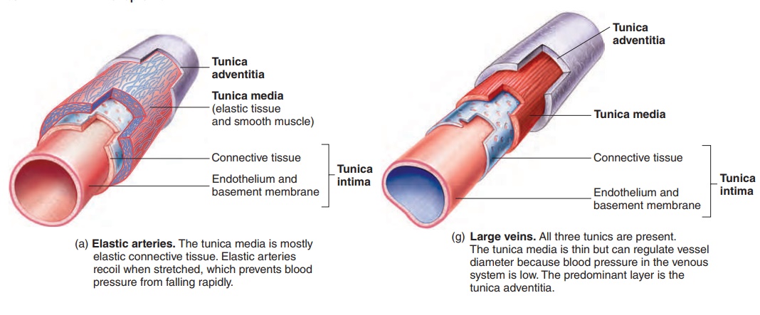
Except in capillaries and venules, blood vessel walls consist of three layers, or tunics (too′ niks). From the inner to the outer wall, the tunics are (1) the tunica intima, (2) the tunica media, and (3) the tunica adventitia, or tunica externa (figure 13.2; see figure 13.1).
The tunica intima (too′ ni-kă in′ ti-mă), or innermost layer, consists of an endothelium composed of simple squamous epithe-lial cells, a basement membrane, and a small amount of connective tissue. In muscular arteries, the tunica intima also contains a layer of thin elastic connective tissue. The tunica media, or middle layer, consists of smooth muscle cells arranged circularly around the blood vessel. It also contains variable amounts of elastic and collagen fibers, depending on the size and type of the vessel. In muscular arteries, a layer of elastic connective tissue forms the outer margin of the tunica media. Thetunica adventitia (ad-ven-tish′ ă) is composed of dense connective tissue adjacent to the tunica media; the tissue becomes loose connective tissue toward the outer portion of the blood vessel wall.
Arteries
Elastic arteries are the largest-diameter arteries and have thethickest walls (see figure 13.1a). Compared to other arteries, a greater proportion of their walls is composed of elastic tissue, and a smaller proportion is smooth muscle. The aorta and pulmonary trunk are examples of elastic arteries. Elastic arteries stretch when the ventricles of the heart pump blood into them. The elastic recoil of these arteries prevents blood pressure from falling rapidly and maintains blood flow while the ventricles are relaxed.
The muscular arteries include medium-sized and small arter-ies. The walls of medium-sized arteries are relatively thick compared to their diameter. Most of the wall’s thickness results from smooth muscle cells of the tunica media (see figure 13.1b). Medium-sized arteries are frequently called distributing arteries because the smooth muscle tissue enables these vessels to control blood flow to different body regions. Contraction of the smooth muscle in blood vessels, called vasoconstriction (vā′ sō-kon-strik′ shŭn), decreases blood vessel diameter and blood flow. Relaxation of the smooth muscle in blood vessels, called vasodilation (vā′ sō-dı̆-lā′ shŭn), increases blood vessel diameter and blood flow.
Medium-sized arteries supply blood to small arteries. Small arteries have about the same structure as the medium-sized arteries, except for a smaller diameter and thinner walls. The smallest of the small arteries have only three or four layers of smooth muscle in their walls.
Arterioles (ar-tēr′ ē-ōlz) transport blood from small arteries tocapillaries. Arterioles (see figure 13.1c) are the smallest arteries in which the three tunics can be identified; the tunica media consists of only one or two layers of circular smooth muscle cells. Small arter-ies and arterioles are adapted for vasodilation and vasoconstriction.
Capillaries
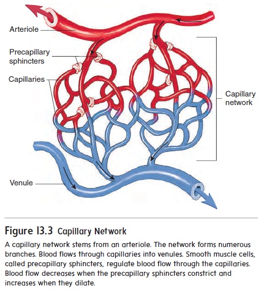
Blood flows from arterioles into capillaries, which branch to form networks (figure 13.3; see figure 13.1d). Blood flow through cap-illaries is regulated by smooth muscle cells called precapillarysphincterslocated at the origin of the branches. Capillary wallsconsist of endothelium (en-dō-thē′ lē-ŭm), which is a layer of simple squamous epithelium surrounded by delicate loose con-nective tissue. The thin walls of capillaries facilitate diffusion between the capillaries and surrounding cells. Each capillary is 0.5–1 millimeter (mm) long. Capillaries branch without changing their diameter, which is approximately the same as the diameter of a red blood cell (7.5 μm).
Red blood cells flow through most capillaries in single file and are frequently folded as they pass through the smaller-diameter capillaries. As blood flows through capillaries, blood gives up O2 and nutrients to the tissue spaces and takes up CO2 and other by-products of metabolism. Capillary networks are more numerous and more extensive in the lungs and in highly metabolic tissues, such as the liver, kidneys, skeletal muscle, and cardiac muscle, than in other tissue types.
Veins
Blood flows from capillaries into venules and from venules into small veins. Venules (ven′ oolz) have a diameter slightly larger than that of capillaries and are composed of endothelium rest-ing on a delicate connective tissue layer (see figure 13.1e). The structure of venules, except for their diameter, is very similar to that of capillaries. Small veins are slightly larger in diameter than venules. All three tunics are present in small veins. The tunica media contains a continuous layer of smooth muscle cells, and the connective tissue of the tunica adventitia surrounds the tunica media (see figure 13.1f ).
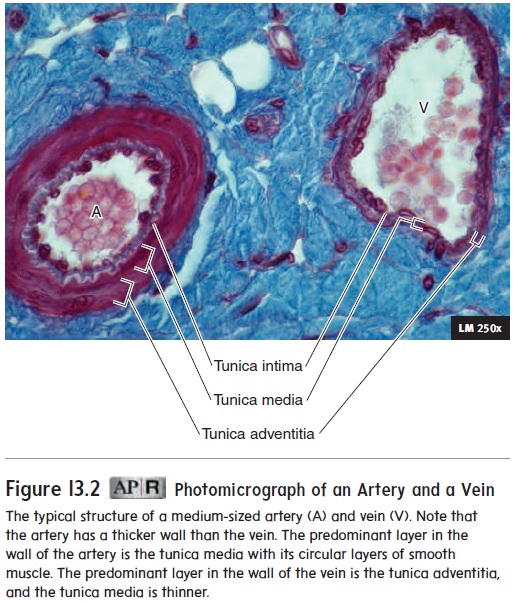
Medium-sized veins collect blood from small veins and deliver it to large veins. The three thin but distinctive tunics make up the wall of the medium-sized and large veins. The tunica media con-tains some circular smooth muscle and sparsely scattered elastic fibers. The predominant layer is the outer tunica adventitia, which consists primarily of dense collagen fibers (see figures 13.1g and 13.2). Consequently, veins are more distensible than arteries. The connective tissue of the tunica adventitia determines the degree to which they can distend.
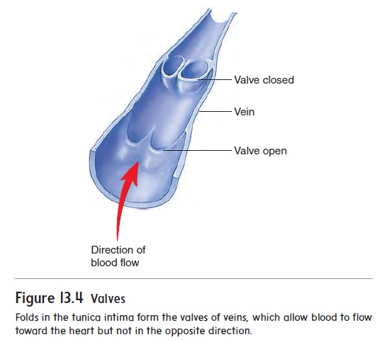
Veins having diameters greater than 2 mm contain valves, which allow blood to flow toward the heart but not in the opposite direction (figure 13.4). Each valve consists of folds in the tunica intima that form two flaps, which are similar in shape and func-tion to the semilunar valves of the heart. There are many valves in medium-sized veins and more valves in veins of the lower limbs than in veins of the upper limbs. This prevents blood from flowing toward the feet in response to the pull of gravity.
Related Topics