Chapter: Medical Physiology: Heart Muscle; The Heart as a Pump and Function of the Heart Valves
Function of the Valves
Function of the Valves
Atrioventricular Valves. TheA-V valves(thetricuspidand mitral valves) prevent backflow of blood from the ventricles to the atria during systole, and the semilu-nar valves (the aortic and pulmonary artery valves)prevent backflow from the aorta and pulmonary ar-teries into the ventricles during diastole. These valves, shown in Figure 9–6 for the left ventricle, close and open passively. That is, they close when a backward pressure gradient pushes blood backward, and they open when a forward pressure gradient forces blood in the forward direction. For anatomical reasons, the thin, filmy A-V valves require almost no backflow to cause closure, whereas the much heavier semilunar valves require rather rapid backflow for a few milliseconds.
Function of the Papillary Muscles. Figure 9–6 alsoshows papillary muscles that attach to the vanes of the A-V valves by the chordae tendineae. The papillary muscles contract when the ventricular walls contract, but contrary to what might be expected, they do nothelp the valves to close. Instead, they pull the vanes of the valves inward toward the ventricles to prevent their bulging too far backward toward the atria during ventricular contraction. If a chorda tendinea becomes ruptured or if one of the papillary muscles becomes paralyzed, the valve bulges far backward during ven-tricular contraction, sometimes so far that it leaks severely and results in severe or even lethal cardiac incapacity.
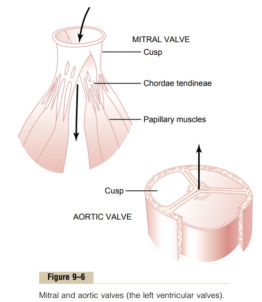
Aortic and Pulmonary Artery Valves. The aortic and pul-monary artery semilunar valves function quite differ-ently from the A-V valves. First, the high pressures in the arteries at the end of systole cause the semilunar valves to snap to the closed position, in contrast to the much softer closure of the A-V valves. Second, because of smaller openings, the velocity of blood ejection through the aortic and pulmonary valves is far greater than that through the much larger A-V valves. Also, because of the rapid closure and rapid ejection, the edges of the aortic and pulmonary valves are subjected to much greater mechanical abrasion than are the A-V valves. Finally, the A-V valves are supported by the chordae tendineae, which is not true for the semi-lunar valves. It is obvious from the anatomy of the aortic and pulmonary valves (as shown for the aortic valve at the bottom of Figure 9–6) that they must be constructed with an especially strong yet very pliable fibrous tissue base to withstand the extra physical stresses.
Related Topics