Chapter: Maternal and Child Health Nursing : Fetal Development, Placenta Development and Fetal Circulation
Fetal and Placental Development and Fetal Circulation
Fetal and Placental Development
and Fetal Circulation
Fertilization
This is
the fusion of the ovum and the spermatozoon, and it initiates the beginning of
a new life. During ovulation the ovum which is released from the ovary is
propelled towards the fallopian tube. During intercourse millions of
spermatozoa released and deposited at the upper vaginal travel towards the
fallopian tube. Aided by the alkaline cervical mucous secretion at the time of
ovulation they travel to the fallopian tubes where they meet the ovum just
released during ovulation. Several hundred of them bind the ovum (zonal
pellucida) but only one spermatozoon can fertilize at a time. The sperm are
viable for 24-72 hours within the female reproductive system. As soon as fusion
occurs between the spermatozoon and the nucleus of the ovum, the zonal
pellucida goes through chemical changes releases an enzyme which make it
impossible for other spermatozoa to penetrate. The membrane of the spermatozoon
breaks; the tail separates and disappears, living a naked male pronucleus. The
fertilized ovum is known as the ZYGOTE. A
new individual has begun its journey till death.
Within a
few hours of fertilization process of rapid mitotic cell division, known as
cleavage starts (2nd cleavage which has been arrested at metaphase
resumes ending in Haploid number). Within 3 days a solid mass of uniform cells
of 16 segments has formed known as the MORULA (Mulberry). During this period
the egg is gently propelled from the ampulla tube where fertilization took
place, along the tube towards the uterine cavity with its peristalsis and
wavelike motion of cilia. With further development central cavity is formed in
the morula and the cell is now moved to one pole, aggregate to form the inner
cell mass from where the embryo and the amnion will be formed, and the fluid on
the other pole. At this stage the ovum is known as the BLASTOCYST, or BLASTULA.(By
4½ days the blastocyst has divided up to 100cells and within approximately 6
days, reaches the decidua of the uterus where implantation takes place.). The
outer wall of the blastocyst is composed of fluid –filled cavity of (
Blastocele surrounded by a single layer of cells) blastocele called TROPHOBLASTfrom where the
placenta and the chorion areformed. It oxidizes the endometrial vascular walls,
it is capable of eating through the decidua and embeds, and it provides the
growing embryo with link to maternal circulation for transportation of
nutrients and Oxygen. At this stage the zonal pellicuda has disappeared and the
Zygote is ready to embed. A layer of cells connect the inner cell mass to the
trophoblast, this forms the body stalk.
Implantation (Embedding): Nidation
Sites
vary from one pregnancy to another, but most often the trophoblast implants
itself in the upper segment of the uterus usually within seven days. Some women
may experience slight vaginal bleeding during implantation (implantation
bleeding). As soon as the trophoblast touches the already prepared uterus
especially with the side of the inner cell mass lies free for 2-3 days, it sticks
to the endometrium and secretes an enzyme which erodes the endometrial cells
and begins to burrow into the endometrium. Three to four days after
implantation, the blastocyst has penetrated very far and completely embedded
and the uterine epithelium covered the entrance the trophoblast has
proliferated and penetrated deeply in the uterus. This takes place by 11th
day after ovulation. The stoma of the endometrium now reacts to the invasion by
accumulating glycogen and lipid and the area become highly vascularised. The
depression formed is filled up with maternal blood which surrounds the ovum. At
this time the endometrium is referred to as the decidua.
Formation of the Decidua
After
fertilization the endometrium of the uterus is known as the decidua. Oestrogen
increases the size to about 4 times of its pre-gravid thickness, the corpus
Luteum, and produce large amount of progesterone which increases the secretion
of the endometrial glands and increases the blood vessels. So it makes the
endometrium to be softer, spongy and vascular for the fertilized ovum to embed
and nourishes itself. The decidua is transformed into 3 layers.
1.
The Basal layer (Basement): This lies immediately
above the myometrium. It remains unchanged in itself but regenerate the new
endometrium after delivery.
2.
The functional layer (cavernous layer): it consists
of tortuous glands rich in secretions. The stoma cells are enlarged in what is
known as the decidua reaction which provides defense against excessive invasion
by the syncytiotrophoblast and limit it to this spongy layer. It provides
anchor for the placenta and allows it to have access to nutrition and 0xygen.
It is the functioning layer.
3.
The compact layer it covers the surface of the
decidua and composed of closely packed stoma cells, polygonal in shape and it
contains necks of glands.
The
blastocyst forms a small nodule in the decidua which bulges out into the
uterine cavity progressively as it continues to enlarge and divides the decidua
into three areas.
1.
Decidua Basalis: This is the area of the decidua
underneath the developing ovum.
2.
Decidua capsularis: the area which covers the ovum.
3.
Decidua
vera (Perietal) (True
Decidua): This lies
in the remainder of the uterine
cavity.
As
development continues the ovum grows and completely fill up the uterine cavity,
at about the 12th week the decidua capsularis comes in contact with
the decidua vera, it fuses with it and degenerates.
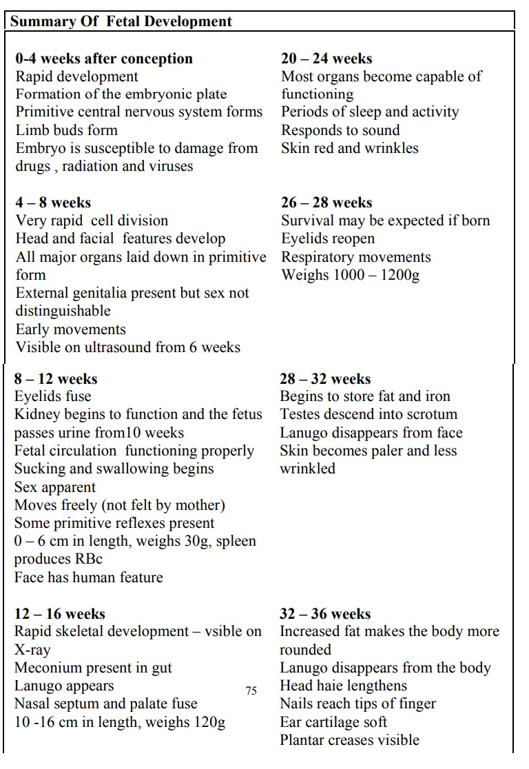
Growth and Development of the Fertilised Ovum
During
the first 8 weeks of pregnancy, embryonic tissues and the surrounding
supportive structures are formed simultaneously. It is during this period that
the embryo is at greatest risk for malformation. From the 8th week
through the end of pregnancy, the embryo is known as the FETUS. The supportive
structures that nourish and maintain the growing fetus are called the fetal
membranes. These include the yolk sac, amnion, chorion, decidua and the
placenta.
The Trophoblast
As
development continues small projections begin to appear all over the surface of
the blastocyst known as the tropoblast, becoming most prolific at the area of
contact – are a of inner cell mass. The trophoblast differentiates into layers.
1.
The outer syncytiotropoblast (syncitium): it is
capable of breaking the decidua tissue during embedding. It erodes the wall of
the blood vessels, making nutrient in the maternal blood accessible to the
developing embryo. It acts as a protective layer between the chorionic villi.
2.
Cytotrophoblast: This is a well defined single
layer of cells which produce Human Chorionic gonadotrophin (HCG). It informs
the corpus Luteum that pregnancy has begun, so as to continue to produce
progesterone and oestrogen. The progesterone maintain the integrity of the
decidua so that shedding does not take place (menstruation is suppressed),
while the high level of oestrogen suppresses the production of FSH. The HCG is
produced in high level in the first trimester and it is the basis for pregnancy
test.
3.
The Mesoderm: Consist of loose connective tissue.
It is continuous with that in the inner cell mass where they join in the body
stalk which later develops into the umbilical cord.
The
trophoblast later form finger like process called –Primitive villi which
develop into placenta and the chorion.
The Inner Cell Mass
As the
trophoblast is developing into the placenta which will nourish the fetus, the
inner cell mass is forming the fetus itself, umbilical cord and the amnion. The
cells differentiate into three layers each of which will form particular parts
of the fetus.
The Ectoderm: Mainly forms the skin, nervous
system,mammary glands salivary glands, Pharynx, nasal passage and crystalline
lens of the eyes, certain lining of the mucosa, hair, nails, and enamel of the
teeth.
The Mesoderm: Forms the bones muscles,
circulatory system oldvessels Reproductory system (ovary and testes), kidneys,
ureters, connective tissues, lymphatic system.
The Endoderm: Lines the yolk sac. It forms the
Alimentary tract,liver, pancreas, lungs, Bladder thyroid glands.
The fetus
develops it’s own blood like other organs in the body. The maternal and the
fetal blood never mix. During the later weeks (4 wks) the organs like the liver
and heart start to function.
The three
layers together are known as the embryonic
plate. Two cavities appear in the inner cell mass one on either sides of
the embryonic plate.
1. The Amniotic Cavity: this lies
on the side of theectoderm. The cavity which is filled with fluid gradually
enlarges and fold round the embryo to enclose it the lining forms the amnion.
It later enlarges in the chronic cavity and comes in contact with the chorionic
membrane.
2. The Yolk
Sac: Lies on the side of the endoderm andprovides nourishment for the embryo
until the placenta(alimentary tract
After
birth the remnants of the yolk sac is the vestigial structure in the base of
the umbilical cord, known as vitelline duct.
The
developing of spring is referred as EMBRYO after fertilization up to 8 weeks
after which the conceptus is known as FETUS until birth.
Related Topics