Chapter: Ophthalmology: The Eyelids
Eyelids: Examination Methods
Examination Methods
The eyelids are examined by direct inspection
under a bright light. A slit lamp may be used for this purpose. Bilateral inspection of the eyelids includes the following aspects:
❖ Eyelid position: Normally the margins of the eyelids are in contact with theeyeball
and the puncta are submerged in the lacus lacrimalis.
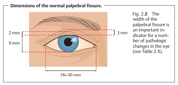
❖ Width of the palpebral fissure: When the eye is open and looking straightahead, the upper lid
should cover the superior margin of the cornea by about 2 mm. Occasionally a
thin strip of sclera will be visible between the cornea and the margin of the
lower lid. The width of the palpebral fissure is normally 6 – 10 mm, and the
distance between the lateral and medial angles of the eye is 28 – 30 mm (Fig.
2.2). Varying widths
of the gaps between the eyelids may be a sign of protrusion of the eyeball,
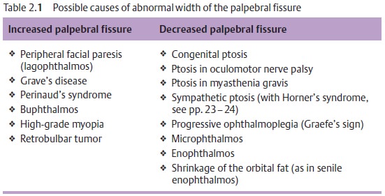
enophthal-mos, or eyeballs of varying size (Table 2.1).
❖ Skin of the eyelid: The skin of the eyelid is thin with only a slight amount ofsubcutaneous fatty tissue. Allergic reaction and inflammation can rapidly cause extensive edema and swelling. In older patients, the skin of the upper eyelid may become increasingly flaccid (cutis laxa senilis). Occa-sionally it can even hang down over the eyelashes and restrict the field of vision (dermatochalasis or blepharochalasis).
Increased palpebral fissure
❖ Peripheral facial paresis (lagophthalmos)
❖ Grave’s disease
❖ Perinaud’s syndrome ❖ Buphthalmos
❖ High-grade myopia ❖ Retrobulbar tumor
Decreased palpebral fissure
❖Congenital ptosis
❖ Ptosis in oculomotor nerve palsy ❖ Ptosis in myasthenia gravis
❖ Sympathetic ptosis (with Horner’s syndrome, )
❖ Progressive ophthalmoplegia (Graefe’s sign) ❖ Microphthalmos
❖ Enophthalmos
❖ Shrinkage of the orbital fat (as in senile
enophthalmos)
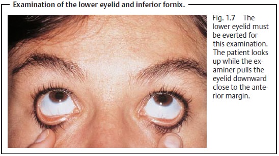
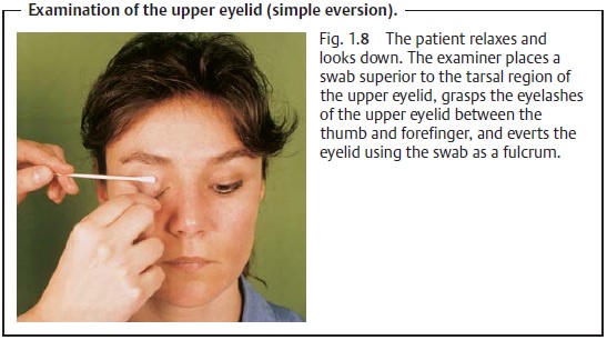
The palpebral conjunctiva is examined by simple eversion of the upper eye-lid (see Figs. 1.7 and 1.8). The normal
palpebral conjunctiva is smooth and shiny without any scar strictures or
papilliform projections.
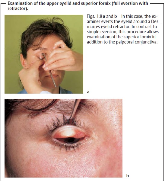
Full eversion of the upper eyelid with a Desmarres eyelid retractor (seeFig. 1.9, p. 9) allows examination of the superior fornix (for normal
appear-ance, see palpebral conjunctiva).
Related Topics