Chapter: Medical Surgical Nursing: Assessment and Management of Patients With Eye and Vision Disorders
Eye - Anatomic and Physiologic Overview
Anatomic and Physiologic
Overview
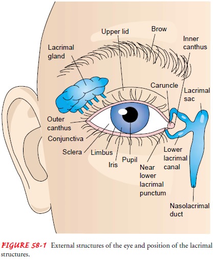
Unlike most organs of the body, the eye is available for external examination, and its anatomy is more easily assessed than many other body parts (Fig. 58-1). The eyeball, or globe, sits in a pro-tective bony structure known as the orbit. Lined with muscle and connective and adipose tissues, the orbit is about 4 cm high, wide, and deep, and it is shaped roughly like a four-sided pyramid, sur-rounded on three sides by the sinuses: ethmoid (medially), frontal (superiorly), and maxillary (inferiorly).
The optic nerve and the ophthalmic
artery enter the orbit at its apex through the optic foramen. The eyeball is
moved though all fields of gaze by the ex-traocular muscles. The four rectus
muscles and two oblique mus-cles (Fig. 58-2) are innervated by cranial nerves (CN)
III, IV, and VI. Normally, the movements of the two eyes are coordinated, and
the brain perceives a single image.

The
eyelids, composed of thin elastic skin that covers striated and smooth muscles,
protect the anterior portion of the eye. The eyelids contain multiple glands,
including sebaceous, sweat, and accessory lacrimal glands, and they are lined
with conjunctival material. The upper lid normally covers the uppermost portion
of the iris and is innervated by the oculomotor nerve (CN III). The lid margins
contain meibomian glands, the inferior and su-perior puncta, and the eyelashes.
The triangular spaces formed by the junction of the eyelids are known as the
inner or medial can-thus and the outer or lateral canthus. With every blink of
the eyes, the lids wash the cornea and conjunctiva with tears.
Tears
are vitally important to eye health. They are formed by the lacrimal gland and
the accessory lacrimal glands. A healthy tear is composed of three layers:
lipoid, aqueous, and mucoid. If there is a defect in the composition of any of
these layers, the integrity of the cornea may be compromised. Tears are
secreted in response to reflex or emotional stimuli.
The
conjunctiva, a mucous membrane, provides a barrier to the external environment
and nourishes the eye. The goblet cells of the conjunctiva secrete lubricating
mucus. The bulbar con-junctiva covers the sclera, whereas the palpebral
conjunctiva lines the inner surface of the upper and lower eyelids. The
junction of the two portions is known as the fornix.
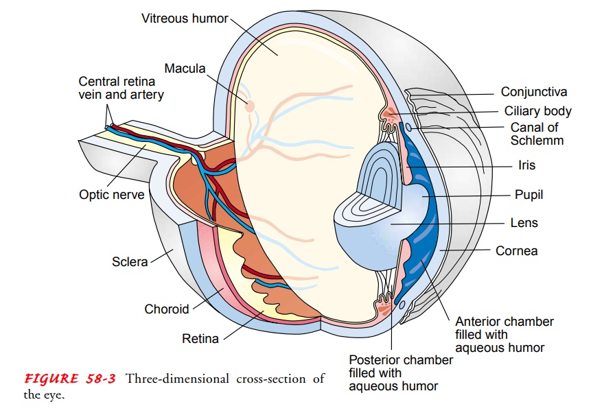
The sclera, commonly known as the white of the eye,
is a dense, fibrous structure that comprises the posterior five sixths of the
eye (Fig. 58-3). The sclera helps to maintain the shape of the eyeball and
protects the intraocular contents from trauma. The sclera may have a slightly
bluish tinge in young children, a dull white color in adults, and a slightly
yellowish color in the elderly. Externally, it is overlaid with conjunctiva,
which is a thin, transparent, mu-cous membrane that contains fine blood
vessels. The conjunctiva meets the cornea at the limbus on the outermost edge of the iris.
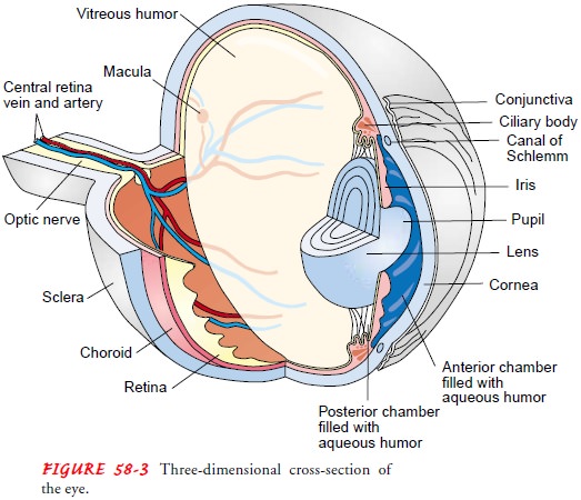
The
cornea (Fig. 58-4), a transparent, avascular, domelike structure, forms the
most anterior portion of the eyeball and is the main refracting surface of the eye.
It is composed of five lay-ers: epithelium, Bowman’s membrane, stroma,
Descemet’s mem-brane, and endothelium. The epithelial cells are capable of
rapid replication and are completely replaced every 7 days.
Behind
the cornea lies the anterior chamber,
filled with a continually replenished supply of clear aqueous humor, which
nourishes the cornea. The aqueous humor is produced by the cil-iary body, and
its production is related to the intraocular pressure (IOP). Normal pressure is
10 to 21 mm Hg.
The uvea consists of the iris, the ciliary body,
and the choroid. The iris, or colored part of the eye, is a highly
vascularized, pig-mented collection of fibers surrounding the pupil. The pupil
is a space that dilates and constricts in response to light. Normal pupils are
round and constrict symmetrically when a bright light shines on them. About 20%
of the population has pupils that are slightly unequal in size but that respond
equally to light. Dilation and constriction are controlled by the sphincter and
dilator pupil-lae muscles. The dilator muscles are controlled by the
sympathetic nervous system, whereas the sphincter muscles are controlled by the
parasympathetic nervous system.
Directly behind the pupil and iris lies the lens, a
colorless and almost completely transparent, biconvex structure held in
posi-tion by zonular fibers. It is avascular and has no nerve or pain fibers.
The lens enables focusing for near vision and refocusing for distance vision.
The ability to focus and refocus is called ac-commodation.
The lens is suspended behind the iris by thezonules and is connected to the
ciliary body. The ciliary body controls accommodation through the zonular
fibers and the cil-iary muscles. The aqueous humor is anterior to the lens;
posterior to the lens is the vitreous
humor. All cells formed throughout life are retained by the lens, which
makes the cell structure of the lens susceptible to the degenerative effects of
aging. The lens con-tinues to grow throughout life, laying down fibers in
concentric rings. This gradual thickening becomes evident in the fifth decade
of life and eventually results in an increasingly dense core or nucleus, which
can limit accommodative powers.
The posterior
chamber is a small space between the vitreous and the iris. Aqueous fluid
is manufactured in the posterior cham-ber by the ciliary body. This aqueous
fluid flows from the poste-rior chamber into the anterior chamber, from which
it drains through the trabecular meshwork into the canal of Schlemm.
The
choroid lies between the retina and the sclera. It is a vas-cular tissue,
supplying blood to the portion of the sensory retina closest to it.
The
ocular fundus is the largest chamber of the eye and con-tains the vitreous humor, a clear, gelatinous
substance, com-posed mostly of water and encapsulated by a hyaloid membrane.
The vitreous humor occupies about two thirds of the eye’s vol-ume and helps
maintain the shape of the eye. As the body ages, the gel-like characteristics
are gradually lost, and various cells and fibers cast shadows that the patient
perceives as “floaters.” The vitreous is in continuous contact with the retina
and is attached to the retina by scattered collagenous filaments. The vitreous
shrinks and shifts with age.
The innermost surface of the fundus is the retina. The retina is composed of 10 microscopic layers and has the consistency of wet tissue paper. It is neural tissue, an extension of the optic nerve. Viewed through the pupil, the landmarks of the retina are the optic disc, the retinal vessels, and the macula. The point of entrance of the optic nerve into the retina is the optic disc. The optic disc is oval or circular, is pink, and has sharp margins.
In the disc, a physiologic depression or cup is present centrally, with the retinal blood vessels emanating from it. The retinal tissues arise from the optic disc and line the inner surface of the vitreous chamber. The retinal vessels also enter the eye through the optic nerve, branching out through the retina and forming superior and inferior arcades. The area of the retina responsible for central vision is the macula. The rest of the retina is responsible for pe-ripheral vision. In the center of the macula is the most sensitive area, the fovea, which is avascular and surrounded by the supe-rior and inferior vascular arcades. Two important layers of the retina are the retinal pigment epithelium (RPE) and the sensory retina. A single layer of cells constitutes the RPE, and these cells have numerous functions, including the absorption of light. The sensory retina contains the photoreceptor cells: rods and cones. Rods and cones are long, narrow cells shaped like rods or cones. The rods are mainly responsible for night vision or vision in low light, whereas the cones provide the best vision for bright light, color vision, and fine detail. Cones are distributed throughout the retina with their greatest concentration in the fovea. Rods are ab-sent in the fovea.
Good visual acuity depends on a healthy,
functioning eyeball and an intact visual pathway (Fig. 58-5). This pathway is
made up of the retina, optic nerve, optic chiasm, optic tracks, lateral
genic-ulate bodies, optic radiations, and the visual cortex area of the brain.
The pathway is an extension of the central nervous system.
The optic nerve is also known as the second cranial nerve (CN II). Its purpose is to transmit impulses from the retina to the occipital lobe of the brain. The optic nerve head, or optic disc, is the physiologic blind spot in each eye. The optic nerve leaves the eye and then meets the optic nerve from the other eye at the optic chiasm.
The chiasm is the anatomic point at which the nasal fibers
from the nasal retina of each eye cross to the opposite side of the brain. The
nerve fibers from the temporal retina of each eye remain uncrossed. Fibers from
the right half of each eye, which would be the left visual field, therefore
carry impulses to the right occipital lobe. Fibers from the left half of each
eye, or the right visual field, carry impulses to the left occipital lobe.
Beyond the chiasm, these fibers are known as the optic tract. The optic tract
continues on to the lateral geniculate body. The lateral geniculate body leads
to the optic radiations and then to the cortex of the occipital lobe of the
brain.
Related Topics