Chapter: Basic Radiology : Radiology of the Chest
Exercise: Mediastinal Masses and Compartments
EXERCISE 4-11.
MEDIASTINAL MASSES AND COMPARTMENTS
4-17. The chest radiograph
in Figure 4-52 shows
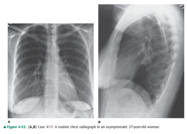
A.
an anterior mediastinal mass.
B.
a middle mediastinal mass.
C.
a posterior mediastinal mass.
D.
a superior mediastinal mass.
4-18. The chest
radiograph in Figure 4-53 shows
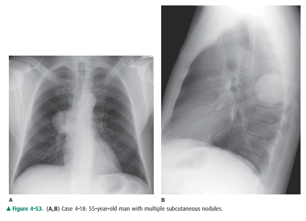
A.
an anterior mediastinal mass.
B.
a middle mediastinal mass.
C.
a posterior mediastinal mass.
D.
a superior mediastinal mass.
4-19. The chest
radiograph in Figure 4-54 shows
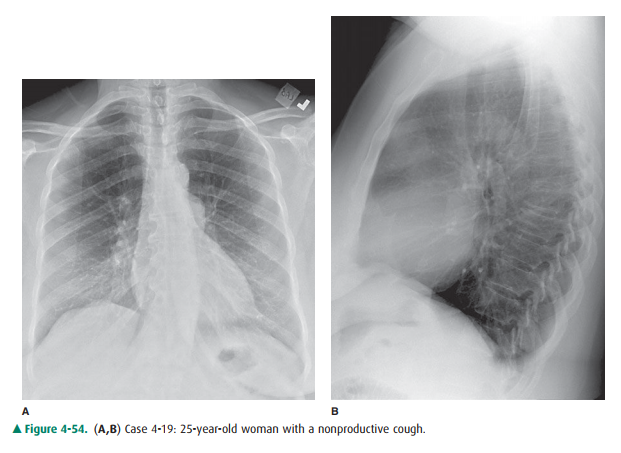
A.
an anterior mediastinal mass.
B.
a middle mediastinal mass.
C.
a posterior mediastinal mass.
D.
a superior mediastinal mass.
Radiologic Findings
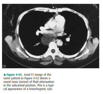
4-17. A spherical mass 4
cm in diameter is present in the subcarinal region on the frontal radiograph
(Figure 4-52 A), and superimposed on the hilar region on the lateral radiograph
(Figure 4-52 B). CT (Figure 4-55) shows that the lesion is of fluid attenuation
(greater attenuation than the subcutaneous fat, but less atten-uation than
muscle). This mass is in the middle mediastinum. (B is the correct answer to
Question 4-17.) In an asymptomatic individual, this most likely represents a
congenital bronchogenic cyst. These masses can grow to sufficient size to cause
symptoms such as dyspnea or dysphagia owing to compression of the trachea or
esophagus. Bron-chogenic cysts may also occur within the lungs and are often
surgically resected because of the likelihood of pulmonary infection. The
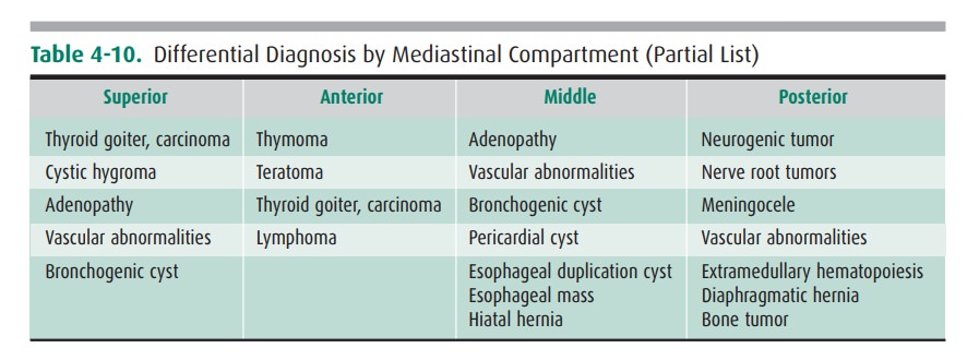
differential diagnosis of a middle mediastinal mass can be seen in Table 4-10.
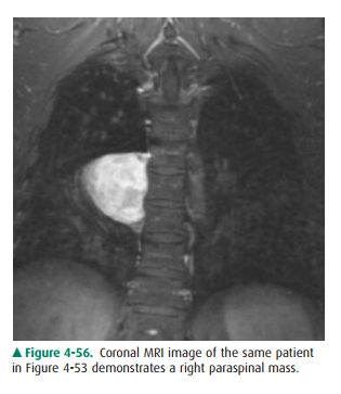
4-18. The frontal
radiograph (Figure 4-53) shows a lobu-lated mass to the right of the lower
thoracic vertebrae. Note that the right heart border remains visible,
sug-gesting that this mass is either anterior or posterior to
Discussion
Two methods of dividing the
mediastinum for radiographic purposes are in common use. The radiographic
divisions are arbitrary and are intended to provide the most appro-priate
differential diagnosis for abnormalities that occur in these locations. Neither
of the divisions follows the divi-sions used by anatomists. In the older
system, the medi-astinum is divided into three compartments. The anterior
mediastinum is that portion of the mediastinum that is an-terior to the
anterior margin of the trachea and along the posterior margin of the
pericardium and inferior vena cava. The posterior mediastinum lies behind a plane
that extends the length of the thorax behind a line drawn 1 cm posteri-orly to
the anterior margin of the vertebral column. The middle mediastinum is the
region between these two boundaries. This system has been superseded by a
four-compartment model, which designates a superior mediasti-nal compartment as
the space that lies above a planeextending from the sternomanubrial junction to
the lower border of the fourth thoracic vertebra. The anterior medi-astinum is
just caudad to the superior compartment and is anterior to a plane extending
along the anterior aspect of the tracheal air column and along the anterior
pericardium. Note that the heart shifts from the anterior to the middle
mediastinum with the four-compartment system. The mid-dle mediastinum occupies
the area from the anterior peri-cardium backward to a plane 1 cm posterior to
the anterior margin of the vertebral column. The addition of the fourth
compartment occurred when CT was developed and it be-came easier to identify
structures in each compartment.
The differential diagnosis of
lesions occurring in each compartment is in part dependent on the structures
that exist there (see Table 4-10). Note that there are vascular structures and
lymph nodes in each of the compartments. Therefore, abnormalities of the blood
vessels (eg, aneurysms) and lymph node diseases (eg, lymphoma) would have to be
included in the differential diagnosis of diseases occurring there. The
differential diagnosis lists include the most com-mon disorders occurring in
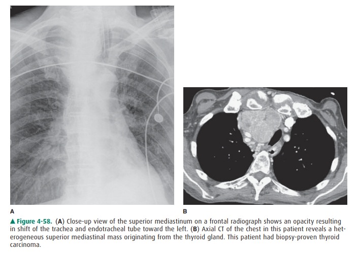
each region. The most com-mon mass to occur in the superior mediastinum is an
enlarged substernal thyroid, which may become large enough to extend into the
anterior or middle mediastinum (Figure 4-58 A,B).
Related Topics