Chapter: Medical Physiology: The Eye: I. Optics of Vision
Errors of Refraction - Optics of the Eye
Errors of Refraction
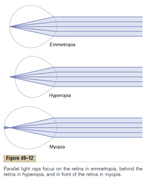
Emmetropia (Normal Vision). As shown in Figure 49–12, theeye is considered to be normal, or “emmetropic,” if par-allel light rays from distant objects are in sharp focus on the retina when the ciliary muscle is completely relaxed.
This means that the emmetropic eye can see all distant objects clearly with its ciliary muscle relaxed. However, to focus objects at close range, the eye must contract its ciliary muscle and thereby provide appropriate degrees of accommodation.
Hyperopia (Farsightedness). Hyperopia, which is alsoknown as “farsightedness,” is usually due to either an eyeball that is too short or, occasionally, a lens system that is too weak. In this condition, as seen in the middle panel of Figure 49–12, parallel light rays are not bent sufficiently by the relaxed lens system to come to focus by the time they reach the retina. To overcome this abnormality, the ciliary muscle must contract to increase the strength of the lens. By using the mechanism of accommodation, a farsighted person is capable of focus-ing distant objects on the retina. If the person has used only a small amount of strength in the ciliary muscle to accommodate for the distant objects, he or she still has
much accommodative power left, and objects closer and closer to the eye can also be focused sharply until the ciliary muscle has contracted to its limit. In old age, when the lens becomes “presbyopic,” a farsighted person is often unable to accommodate the lens suffi-ciently to focus even distant objects, much less near objects.
Myopia (Nearsightedness). In myopia, or“nearsighted-ness,” when the ciliary muscle is completely relaxed, the light rays coming from distant objects are focused in front of the retina, as shown in the bottom panel of Figure 49–12. This is usually due to too long an eyeball, but it can result from too much refractive power in the lens system of the eye.
No mechanism exists by which the eye can decrease the strength of its lens to less than that which exists when the ciliary muscle is completely relaxed. A myopic person has no mechanism by which to focus distant objects sharply on the retina. However, as an object moves nearer to the person’s eye, it finally gets close enough that its image can be focused. Then, when the object comes still closer to the eye, the person can use the mechanism of accommodation to keep the image focused clearly. A myopic person has a definite limiting “far point” for clear vision.
Correction of Myopia and Hyperopia by Use of Lenses.
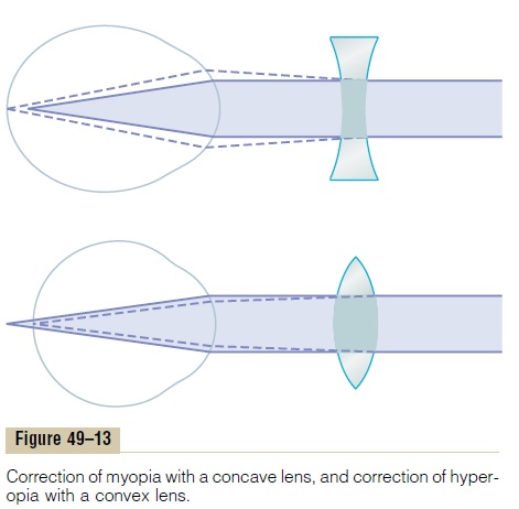
It will be recalled that light rays passing through a concave lens diverge. If the refractive surfaces of the eye have too much refractive power, as in myopia, this excessive refractive power can be neutralized by placing in front of the eye a concave spherical lens, which will diverge rays. Such correction is demonstrated in the upper diagram of Figure 49–13.
Conversely, in a person who has hyperopia—that is, someone who has too weak a lens system—the abnor-mal vision can be corrected by adding refractive power using a convex lens in front of the eye. This correction is demonstrated in the lower diagram of Figure 49–13.
One usually determines the strength of the concave or convex lens needed for clear vision by “trial and error”—that is, by trying first a strong lens and then a stronger or weaker lens until the one that gives the best visual acuity is found.
Astigmatism
Astigmatism is a refractive error of the eye that causes the visual image in one plane to focus at a different dis-tance from that of the plane at right angles. This most often results from too great a curvature of the cornea in one plane of the eye. An example of an astigmatic lens would be a lens surface like that of an egg lying sidewise to the incoming light. The degree of curvature in the plane through the long axis of the egg is not nearly as great as the degree of curvature in the plane through the short axis.
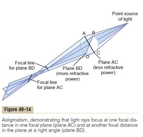
Because the curvature of the astigmatic lens along one plane is less than the curvature along the other plane, light rays striking the peripheral portions of the lens in one plane are not bent nearly as much as the rays striking the peripheral portions of the other plane. This is demonstrated in Figure 49–14, which shows rays of light originating from a point source and passing through an oblong, astigmatic lens. The light rays in the vertical plane, indicated by plane BD, are refracted greatly by the astigmatic lens because of the greater cur-vature in the vertical direction than in the horizontal direction. By contrast, the light rays in the horizontal plane, indicated by plane AC, are not bent nearly as much as the light rays in vertical plane BD. It is obvious that light rays passing through an astigmatic lens do not all come to a common focal point, because the light rays passing through one plane focus far in front of those passing through the other plane.
The accommodative power of the eye can never compensate for astigmatism because, during accommo-dation, the curvature of the eye lens changes approxi-mately equally in both planes; therefore, in astigmatism, each of the two planes requires a different degree of accommodation. Thus, without the aid of glasses, a person with astigmatism never sees in sharp focus.
Correction of Astigmatism with a Cylindrical Lens. Onemay consider an astigmatic eye as having a lens system made up of two cylindrical lenses of different strengths and placed at right angles to each other. To correct for astigmatism, the usual procedure is to find a spherical lens by trial and error that corrects the focus in one of the two planes of the astigmatic lens. Then an additional cylindrical lens is used to correct the remaining error in the remaining plane. To do this, both the axis and the strength of the required cylindrical lens must be determined.
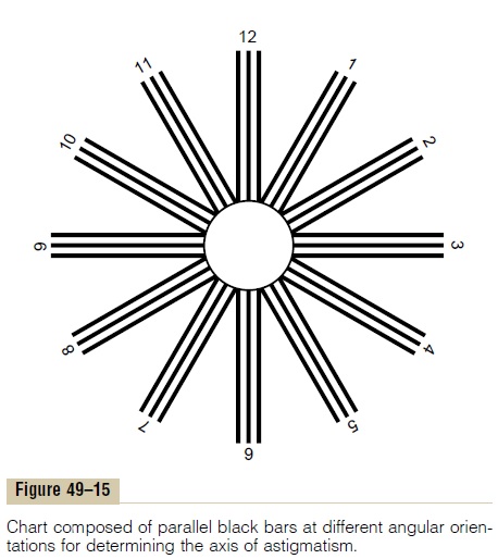
There are several methods for determining the axis of the abnormal cylindrical component of the lens system of an eye. One of these methods is based on the use of parallel black bars of the type shown in Figure 49–15. Some of these parallel bars are vertical, some horizontal, and some at various angles to the vertical and horizontal axes. After placing various spherical lenses in front of the astigmatic eye, a strength of lens is usually found that causes sharp focus of one set of parallel bars but does not correct the fuzziness of the set of bars at right angles to the sharp bars. It can be shown from the physical principles of optics discussed earlier that the axis of the out-of-focuscylindrical component of the optical system is parallel to the bars that are fuzzy. Once this axis is found, the examiner tries progressively stronger and weaker posi-tive or negative cylindrical lenses, the axes of which are placed in line with the out-of-focus bars, until the patient sees all the crossed bars with equal clarity. When this has been accomplished, the examiner directs the optician to grind a special lens combining both the spherical correction and the cylindrical correction at the appropriate axis.
Correction of Optical Abnormalities by Use of Contact Lenses
Glass or plastic contact lenses can be inserted that fit snugly against the anterior surface of the cornea. These lenses are held in place by a thin layer of tear fluid that fills the space between the contact lens and the anterior eye surface.
A special feature of the contact lens is that it nullifies almost entirely the refraction that normally occurs at the anterior surface of the cornea. The reason for this is that the tears between the contact lens and the cornea have a refractive index almost equal to that of the cornea, so that the anterior surface of the cornea no longer plays a significant role in the eye’s optical system. Instead, the outer surface of the contact lens plays the major role. Thus, the refraction of this surface of the contact lens substitutes for the cornea’s usual refraction. This is especially important in people whose eye refrac-tive errors are caused by an abnormally shaped cornea, such as those who have an odd-shaped, bulging cornea—a condition called keratoconus. Without the contact lens, the bulging cornea causes such severe abnormality of vision that almost no glasses can correct the vision satisfactorily; when a contact lens is used, however, the corneal refraction is neutralized, and normal refraction by the outer surface of the contact lens is substituted.
The contact lens has several other advantages as well, including (1) the lens turns with the eye and gives a broader field of clear vision than glasses do, and (2) the contact lens has little effect on the size of the object the person sees through the lens, whereas lenses placed 1 centimeter or so in front of the eye do affect the size of the image, in addition to correcting the focus.
Cataracts
“Cataracts” are an especially common eye abnormality that occurs mainly in older people. A cataract is a cloudy or opaque area or areas in the lens. In the early stage of cataract formation, the proteins in some of the lens fibers become denatured. Later, these same proteins coagulate to form opaque areas in place of the normal transparent protein fibers.
When a cataract has obscured light transmission so greatly that it seriously impairs vision, the condition can be corrected by surgical removal of the lens. When this is done, the eye loses a large portion of its refractive power, which must be replaced by a powerful convex lens in front of the eye; usually, however, an artificial plastic lens is implanted in the eye in place of the removed lens.
Related Topics