Chapter: Human Neuroanatomy(Fundamental and Clinical): Tracts of Spinal Cord and Brainstem
Disorders of Equilibrium - Tracts of Spinal Cord and Brainstem
Disorders of Equilibrium
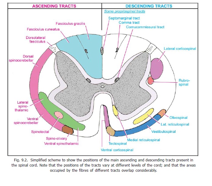
We have seen that the maintenance of equilibrium, and of correct posture, is dependent on reflex arcs involving various centres including the spinal cord, the cerebellum, and the vestibular nuclei. Afferent impulses for these reflexes are carried by the posterior column tracts (fasciculus gracilis and fasciculus cuneatus), the spinocerebellar tracts, and others. Efferents reach neurons of the ventral grey column (anterior horn cells) through rubrospinal, vestibulospinal, and other ‘extrapyramidal’ tracts. Interruption of any of these pathways; or lesions in the cerebellum, the vestibular nuclei and other centres concerned; can result in various abnormalities involving maintenance of posture and coordination of movements.
Inability to maintain the equilibrium of the body, while standing, or while walking, is referred to as ataxia. This may occur as a result of the interruption of afferent proprioceptive pathways (sensory ataxia). Disease of the cerebellum itself, or of efferent pathways, results in more severe disability.Lack of proprioceptive information can be compensated to a considerable extent by information received through the eyes. The defects are, therefore, much more pronounced with the eyes closed (Romberg’ssign)..
Propriospinal tracts
In addition to the various ascending and descending tracts described in preceding sections, the white matter of the spinal cord contains numerous fibres that interconnect neurons in different segments of the cord. These fibres, that are both ascending and descending, constitute the propriospinal or intersegmental tracts. These tracts are present in the anterior, lateral and posterior funiculi and occupy areas adjacent to spinal grey matter. In addition to intersegmental fibres this region is also believed to contain some reticulospinal and descending autonomic fibres.
Nerve fibres in propriospinal tracts are classified as long, intermediate or short. Neurons from which they arise lie in laminae V to VIII. They are interneurons, but can carry impulses arising in one segment to distant regions. Some propriospinal tracts that have been given special names are as follows.
1. Thedorsolateral fasciculus(or tract of Lissauer) contains many propriospinal fibres. It alsocontains ascending and descending branches of axons entering the spinal cord through dorsal nerve roots. Many fibres are axons of cells in the substantia gelatinosa.
2. Several relatively discrete bundles of fibres have been described in the dorsal funiculi. These arethe comma tract (also called the semilunar tract or interfascicular fasciculus); the septomarginal tract and the cornu commissural tract (Fig. 9.2).
Lamination of fibres in tracts
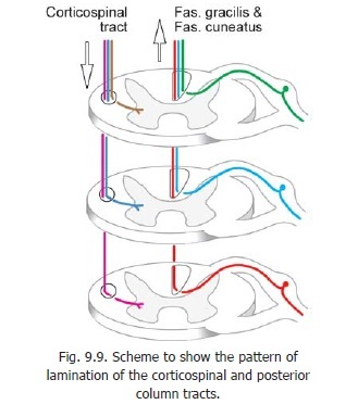
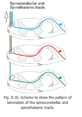
An interesting aspect of the larger tracts of the spinal cord is that the fibres entering or leaving the tracts at various levels of the cord appear to be arranged in layers. This is spoken of as lamination. Lamination in individual tracts is easily remembered once the reason for it is understood. From Fig. 9.9 (left half) it will be clear that in the case of any descending tract located lateral to the grey matter in which the fibres of the tract terminate, the fibres for the lowest segments of the cord must be placed most laterally if intercrossing of fibres is to be avoided. This explains the arrangement seen in the lateral corticospinal tract. In the case of an ascending tract the arrangement of fibres depends on whether the incoming fibres approach the tract from the medial or lateral side. In the case of the posterior column tracts the fibres (derived from the dorsal nerve roots) enter from the lateral side. Hence as shown in Fig. 9.9 (right half) the lowest fibres are situated most medially. On the contrary in the case of spinothalamic and spinocerebellar tracts, the fibres arise in the spinal grey matter and enter the tracts from the medial side. Hence, as shown in Fig. 9.10 the lowest fibres are most lateral.
Control of conduction through ascending tracts
There are observations to show that conduction through ascending tracts can be controlled (inhibited or facilitated) under the influence of axons descending from higher centres to the neurons which give origin to the tracts.
Stimulation of certain regions in the brainstem reticular formation inhibits transmission of pain through spinothalamic tracts. The centres of the reticular formation concerned are said to constitute and endogenous analgesic system.
Transmission of painful impulses can also be inhibited by stimulation of areas of cerebral hemisphere involved in transmission or perception of the same (i.e., thalamus, parts of cerebral cortex). Conversely, stimulation of some other areas can increase transmission of painful stimuli.
Corticospinal fibres also inhibit neurons giving rise to spinocerebellar tracts. On the other hand vestibulospinal and rubrospinal fibres stimulate the neurons.
Control of conduction of different impulses is believed to be produced mainly by presynaptic inhibition in spinal grey matter. A gate mechanism that determines whether noxious stimuli should be allowed to pass has been postulated and it has been suggested that the substantia gelatinosa may play an important role in this regard. Although the existence of some kind of gate mechanism is accepted, its exact nature, and the role of the substantia gelatinosa are not established.
Related Topics