Chapter: The Massage Connection ANATOMY AND PHYSIOLOGY : Nervous System
Cranial Nerves
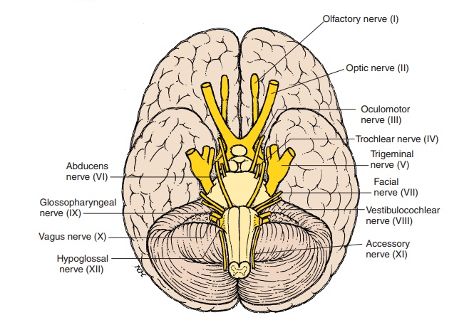
Cranial Nerves
Cranial nerves are part of the peripheral nervous system, which is connected to the brain. Twelve pair of cranial nerves arise from the ventrolateral aspect of the brain (Figure 5.37E). The cranial nerves are num-bered according to their position in the longitudinal axis of the brain, beginning at the cerebrum. Each nerve is named, the name being related to the ap-pearance or function. Similar to the spinal nerves, the cranial nerves may carry sensory fibers, motor fibers, or both; some carry fibers with autonomic function.
The sensory nerves synapse at the brainstem or join the ascending sensory tracts from the rest of the body to reach the thalamus and cerebral cortex. In addition to sensory nerves that carry sensations such as pain, temperature, touch-pressure, some cranial nerves carry impulses generated by special sense or- gans located in the eye (vision), ear (hearing and bal-ance), nose (smell), and tongue (taste).
The motor nerves, such as those in the spinal cord, have numerous synapses and serve as the final com-mon pathway for muscles of the head and neck. The blood vessels, glands, and smooth muscles of the eye (for dilation of pupils) are supplied partly by the au-tonomic component of some of the cranial nerves.
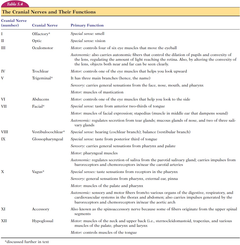
The nerves and their functions are listed in Table 5.4, and the nerves relevant to massage are discussed further.
OLFACTORY NERVE (CRANIAL NERVE I)
Sense of smell and taste are considered visceral sen-sations because they are related to gastrointestinal functions. The flavor of most food is a result of a combination of taste and smell; a person with a cold often complains of diminished taste as a result of a depression of the sense of smell. The receptors for both smell and taste are chemoreceptors (i.e., they are stimulated by chemicals). However, the sense of smell is different in that it is the only sensation that does not relay to the thalamus. Also, there is no direct communication with the sensory cortex.
The Olfactory Mucous Membrane
The receptors for smell are located in the nasal mu-cosa, in a small area about 5 cm2 (0.8 in2) in the roof of the nasal cavity and upper portion of the nasal sep-tum (see Figure 5.40). There are about 10–20 million receptor cells here. Each receptor is the end of a neu-ron; this region is obviously the closest the nervous system gets to the external world. The axons of the neurons go through the cribriform plate of the eth-moid bone and enter the paired projections of the brain—the olfactory bulb and tract. They synapse at the olfactory bulb from which other neurons convey the impulses to the olfactory cortex.
The olfactory cortex consists of two major areas of the brain—the medial olfactory area and the lateral olfactory area. The medial area lies in the middle of the brain, superior and anterior to the hypothalamus and is responsible for reflexes such as licking the lips
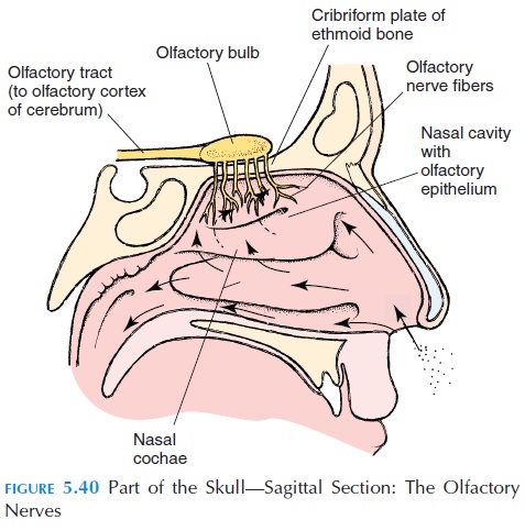
and salivation that occur with response to the smell of food. The lateral olfactory area is located in the base of the brain, spreading to the temporal lobe, and is part of the limbic system with its many connec-tions. Connections with the cortex of the opposite side allow for transfer of memories from one side to the other. Other neurons have connections with the frontal lobe via the thalamus and help with conscious perception and discrimination of smell. The recogni-tion of delectable and detestable food, based on past experience, is a function of the lateral olfactory area. Because of the wide connections, smell has the abil-ity to trigger memories; strong odors can trigger a seizure in individuals with epilepsy.
Stimulation of Olfactory Receptors
Olfactory receptors are stimulated by substances dis-solved in the mucus covering the nasal epithelium. The substance binds to the receptor, which then opens sodium channels, with resultant depolariza-tion and initiation of action potentials. Small sub-stances, with 3–20 carbon atoms; volatile substances (substances that evaporate easily); and those rela-tively soluble in water and lipids have strong odors that are easily smelled. We can distinguish between 2,000 to 4,000 odors; the physiologic basis is not fully known. These receptors adapt quickly (i.e., the per-ception of the odor decreases with time). This is ben-eficial, especially when one is caught in the midst of disagreeable odors! The direction of the smell is iden-tified by the slight difference in the time the smell stimulates the receptors on each side.
In many species of animals, there is a close rela-tionship between smell and sexual function. In many species, behavioral and other physiologic changes are produced in animals of the opposite sex by air-trans-ported hormones. These hormones, known as pheromones; certain fatty acids present in largeamounts at ovulation in female vaginal secretions have been identified as pheromones.
The nose is innervated by the trigeminal nerve, which carries general sensations (e.g., touch, pres-sure, pain, temperature) to the brain. They may be stimulated by irritating odors, and the characteristic smell of such substances as peppermint, menthol, and chlorine is partly a result of the stimulation of pain fibers carried by the trigeminal nerve.
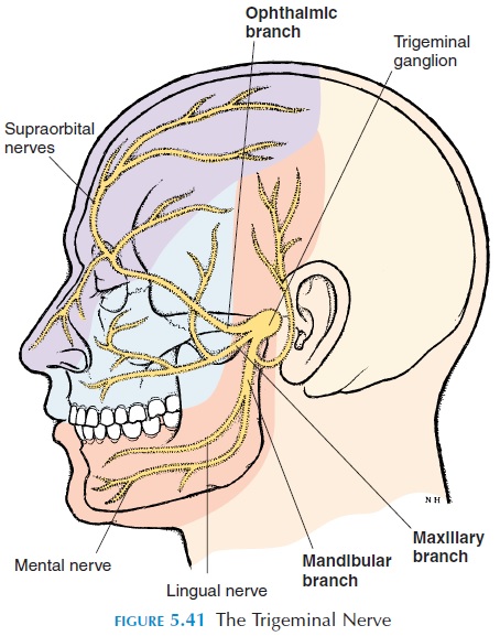
TRIGEMINAL NERVE (CRANIAL NERVE V)
The touch, pressure, and other sensations triggered during a facial massage are carried by the trigeminal nerve. This nerve, as the name suggests, has three major branches (see Figure 5.41). The ophthalmicbranch carries sensations from the eye, nasal cavity,skin on the forehead, upper eyelid, eyebrow, and part of the nose. The maxillary branch carries sensations from the lower eyelid, upper lip, gums, teeth, cheek, nose, palate, and part of the pharynx. The mandibu-lar branch carries sensations from the lower gums,teeth, lips, palate, and part of the tongue. In addition, this nerve controls the muscles of mastication: the temporalis, the masseter, and the pterygoids. The sensory impulses ultimately reach the facial area of the sensory cortex, and the motor reach the respective area of the motor cortex.
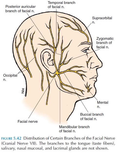
FACIAL NERVE (CRANIAL NERVE VII)
The facial nerve (see Figure 5.42) arises from the pons and contains nerves fibers that have many different functions (Table 5.4). The major motor function of the facial nerve is control of the muscles of facial expres-sion. When the nerve is affected, the muscles of the face become weak, with sagging eyelids; difficulty clos-ing the eyelids tightly, pursing the lips, and blowing out the cheeks; and drooping of the side of the mouth.
Taste Sensations
The taste receptors are located in the walls of tiny projections (papillae) in the tongue and the mucosa of the epiglottis, palate, and pharynx. Similar to smell, these are chemoreceptors, stimulated by sub-stances dissolved in the saliva. Specialized cells, the taste buds,surround the receptors. About 50 nervesinnervate each taste bud, and there are about 10,000 taste buds.
The facial nerve carries taste sensations from the anterior two-thirds of the tongue, the glossopharyn-geal from the posterior one-third, and the vagus nerve from the other areas. From the medulla, the neurons cross over to the other side and reach the cerebral cortex via the thalamus.
In humans, there are four basic tastes: sweet, sour, bitter, and salt. Bitter taste is best sensed in the back of the tongue, sour along the edges, sweet at the tip, and salt on the dorsum, anteriorly. In general, acidic substances taste sour, those containing sodium ions taste salty, and most sweet substances are organic.
VESTIBULOCOCHLEAR NERVE(CRANIAL NERVE VIII)
This nerve has two major divisions: the vestibularnerve and the cochlear nerve. The vestibular nerveconveys sensations from the vestibular apparatus. This organ is stimulated by lin-ear and rotational accelerations of the body and is re-sponsible for equilibrium and balance. After the nerve reaches the medulla, it has extensive connections with the cerebellum. Its connections with cranial nerves III, IV, and VI help the body adjust eye movements ac-cording to the position of the body. Its other connec-tions help the body increase or decrease the tone of different muscle groups to maintain balance.
The cochlear branch carries hearing sensations. The cochlea has many receptors, each stimulated by a specific wavelength. In this way, the pitch of the sound is detected. The intensity of the sound is de-termined by the number of action potentials pro-duced in each receptor. The neurons from the cochlea synapse with others in the medulla, and the impulses ultimately reach the temporal lobe where sound is in-terpreted. Similar to the representation of the body in the primary sensory and motor cortex, there is a rep-resentation in the temporal lobe for various tones.
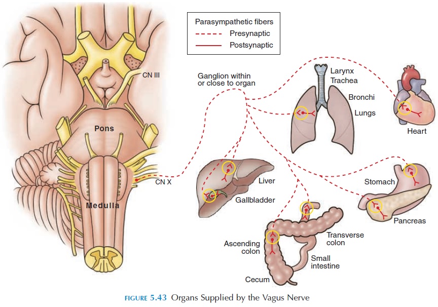
VAGUS (CRANIAL NERVE X)
This nerve has an extensive distribution and is the primary parasympathetic nerve that supplies most of the viscera in the thorax and abdomen (see Figure 5.43). Because it also controls the muscles of the lar-ynx and pharynx, lesions in this nerve can result in hoarseness, difficulty swallowing, and regurgitation of food through the nose..
Related Topics