Chapter: Medical Physiology: The Eye: III. Central Neurophysiology of Vision
Control of Pupillary Diameter
Control of Pupillary Diameter
Stimulation of the parasympathetic nerves also excites the pupillary sphincter muscle, thereby decreasing the pupillary aperture; this is called miosis. Conversely, stimulation of the sympathetic nerves excites the radial fibers of the iris and causes pupillary dilation, called mydriasis.
Pupillary Light Reflex. When light is shone into the eyes,the pupils constrict, a reaction called the pupillary lightreflex. The neuronal pathway for this reflex is demon-strated by the upper two black traces in Figure 51–11. When light impinges on the retina, a few of the result-ing impulses pass from the optic nerves to the pretec-tal nuclei. From here, secondary impulses pass to the Edinger-Westphal nucleus and, finally, back through parasympathetic nerves to constrict the sphincter of theiris. Conversely, in darkness, the reflex becomes inhib-ited, which results in dilation of the pupil.
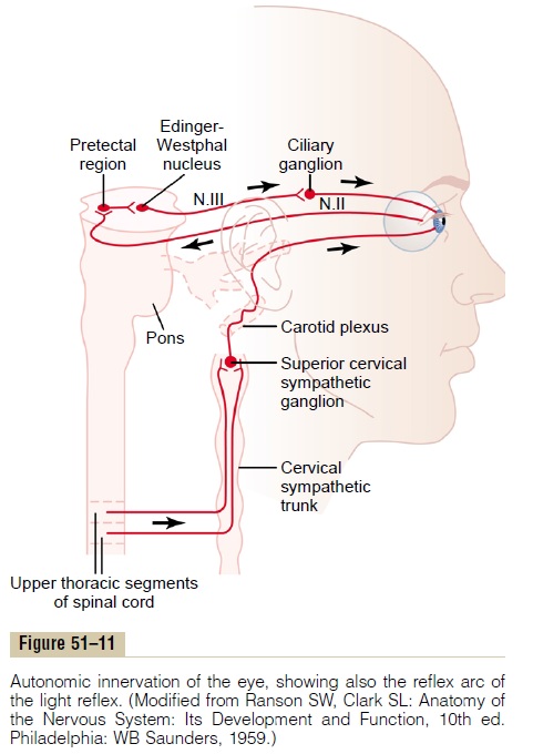
The function of the light reflex is to help the eye adapt extremely rapidly to changing light conditions. The limits of pupillary diameter are about 1.5 millimeters on the small side and 8 millimeters on the large side. Therefore, because light brightness on the retina increases with the square of pupillary diameter, the range of light and dark adap-tation that can be brought about by the pupillary reflex is about 30 to 1—that is, up to as much as 30 times change in the amount of light entering the eye.
Pupillary Reflexes or Reactions in Central NervousSystem Disease. A few central nervous system diseases damagenerve transmission of visual signals from the retinas to the Edinger-Westphal nucleus, thus sometimes blocking the pupillary reflexes. Such blocks frequently occur as a result of central nervous system syphilis, alcoholism,encephalitis, and so forth.The block usually occurs in thepretectal region of the brain stem, although it can result from destruction of some small fibers in the optic nerves.
The final nerve fibers in the pathway through the pre-tectal area to the Edinger-Westphal nucleus are mostly of the inhibitory type. When their inhibitory effect is lost, the nucleus becomes chronically active, causing the pupils to remain mostly constricted, in addition to their failure to respond to light.
Yet the pupils can constrict a little more if the Edinger-Westphal nucleus is stimulated through some other pathway. For instance, when the eyes fixate on a near object, the signals that cause accommodation of the lens and those that cause convergence of the two eyes cause a mild degree of pupillary constriction at the same time. This is called the pupillary reaction to accommodation. A pupil that fails to respond to lightbut does respond to accommodation and is also very small (an Argyll Robertson pupil) is an important diag-nostic sign of central nervous system disease—often syphilis.
Horner’s Syndrome. The sympathetic nerves to the eye areoccasionally interrupted. Interruption frequently occurs in the cervical sympathetic chain. This causes the clini-cal condition called Horner’s syndrome, which consists of the following effects: First, because of interruption of sympathetic nerve fibers to the pupillary dilator muscle, the pupil remains persistently constricted to a smaller diameter than the pupil of the opposite eye. Second, the superior eyelid droops because it is normally main-tained in an open position during waking hours partly by contraction of smooth muscle fibers embedded in the superior eyelid and innervated by the sympathetics. Therefore, destruction of the sympathetic nerves makes it impossible to open the superior eyelid as widely as normally. Third, the blood vessels on the corresponding side of the face and head become persistently dilated. Fourth, sweating (which requires sympathetic nerve signals) cannot occur on the side of the face and head affected by Horner’s syndrome.
Related Topics