Chapter: Human Neuroanatomy(Fundamental and Clinical): Internal Structure of Cerebellum
Connections of Cerebellar Nuclei: Afferents and Efferents
CONNECTIONS OF CEREBELLAR NUCLEI
In the preceding paragraphs mention has been made of many connections involving the cerebellar nuclei. We will now review the connections of each nucleus. The afferents to the nuclei follow a common pattern and will be considered together, but the efferents of each nucleus will be taken up separately.
Afferents to Cerebellar Nuclei
a.Afferents from cerebellar cortex
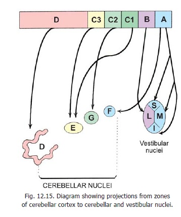
The cerebellar nuclei receive the axons of Purkinje fibres. The zones of cortex from which each nucleus receives fibres are shown in Fig. 12.15. Note that in this figure projections to vestibular nuclei are also shown. These nuclei receive direct fibres from the cortex and are regarded as displaced cerebellar nuclei.
Zone A of the cerebellar cortex projects to the fastigial nucleus, and to vestibular nuclei (other than lateral). The projection to the vestibular nuclei is from the nodule, flocculus and part of the uvula. Fibres from the folium and tuber which are concerned with saccadic eye movements also reach vestibular nuclei.
Zone B projects to the lateral vestibular nucleus.Zones C1 and C3 project to the emboliform nucleus.Zone C2 projects to the globose nucleus. The greater part of the cerebellar cortex (Zone D) projects to the dentate nucleus only.
b.Collaterals of climbing fibres (olivocerebellar fibres)
The inferior olivary complex projects to all parts of the cerebellar cortex. Details of these connections have been summarised in Fig. 12.12. Note that climbing fibres meant for a given zone of cortex give collaterals to the cerebellar nucleus receiving cortical fibres from the same zone. For example:
1.The main inferior olivary nucleus projects to zone D.
2.These fibres give collaterals to the dentate nucleus.
3.Zone D projects to the dentate nucleus.
c.Collaterals of mossy fibres
It may be recalled that all afferents to the cerebellum other than olivocerebellar end as mossy fibres. These fibres give collaterals to cerebellar nuclei.
Reticulocerebellar fibres and spino-cerebellar fibres give collaterals to the fastigial nucleus, the emboliform nucleus and the globose nucleus. Some reticulocerebellar fibres from the reticular nucleus of the pontine tegmentum give collaterals to the dentate nucleus.
Collaterals from pontocerebellar, cuneo-cerebellar and vestibulocerebellar fibres are traditionally described, but their presence is controversial. If present they are few in number.
Efferents of Cerebellar Nuclei
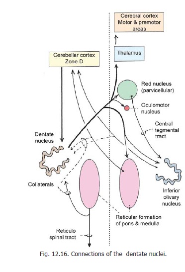
Efferents of dentate nucleus
Efferent fibres arising in the dentate nucleus constitute the greater part of the superior cerebellar peduncle (Fig. 12.16). On reaching the lower part of the midbrain most fibres cross the middle line (in the decussation of the peduncle) and divide into ascending and descending branches.
Most of the ascending fibres reach the thalamus, but some end in the red nucleus. Some ascending fibres end in the nucleus of the oculomotor nerve, and some in the tectum. The descending fibres end in the inferior olivary complex and in the reticular formation.
Some fibres of the superior cerebellar peduncle do not cross. Like the crossed fibres, the uncrossed fibres divide into ascending and descending branches all of which end in the reticular formation.
Efferents of the emboliform nucleus
To understand the connections of the emboliform nucleus think of it as a centre on pathways through which the cerebellar cortex controls motor neurons in the spinal cord. We have seen above that the emboliform nucleus receives afferents from the pars intermedia (paravermal area) of the cerebellar cortex (zones C1, C3). The efferents of the nucleus are as follows (Fig. 12.17).
Fibres arising in the emboliform nucleus pass through the superior cerebellar peduncle. They decussate and divide into ascending and descending branches.
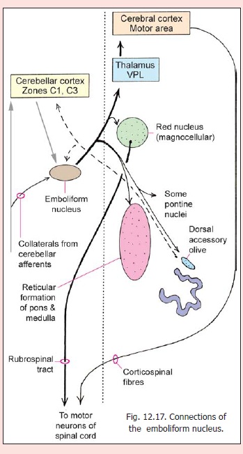
Some ascending fibres end in the red nucleus (magnocellular part). The emboliform nucleus can, therefore, influence motor neurons in the spinal cord through the rubrospinal tract. Other ascending fibres reach the ventral lateral nucleus of the thalamus (posterior part). From here they are relayed to the motor cortex. The cerebellum is, therefore, able to influence motor neurons through the corticospinal tracts also.
Some descending fibres end in the reticular formation (medial part, mainly the reticular nucleus of the pontine tegmentum). These fibres act on spinal neurons through reticulospinal fibres. Other descending fibres end in relation to pontine nuclei. They may influence activity of the ponto-cerebellar projection. Yet other descending fibres end in the (rostral half of the) dorsal accessory olivary nucleus (nucleo-olivary fibres).
Efferents of globose nucleus
Like those from the dentate and emboliform nuclei efferents from the globose nucleus pass through the superior cerebellar peduncle to reach the red nucleus and the thalamus (Fig. 12.18). Some fibres reach the superior colliculus, the central grey matter of the midbrain, raphe nuclei (in the reticular formation), and the nucleus of Darkschewitsch. The nucleus of Darkschewitsch projects to the upper part of the medial accessory olive through the medial tegmental tract.
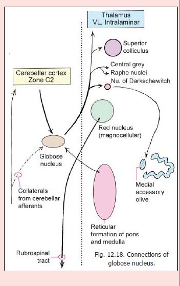
Fibres passing into the descending division of the superior cerebellar peduncle connect the globose nucleus to the reticular nucleus of the pontine tegmentum. Spinal motor neurons can be influenced through this nucleus.
Connections of the fastigial nucleus
A general idea of the afferents to this nucleus has been given. We will now consider further details of both its afferents and efferents (Fig. 12.19). It is easier to understand the numerous connections of the fastigial nucleus if we consider them in relation to the functions they serve.
A.Control of muscle action (axial andproximal limb muscles) in response to labyrinthine stimuli.
1. Fibres of the vestibular nerve carrying labyrinthine impulses project to zone A of the cerebellar cortex. (The area includes the medial part of vermis, whole of posterior vermis and flocculus. The area corresponds roughly to the vestibulocerebellum of earlier descriptions).These fibres may reach the cortex directly, or after relay in vestibular nuclei.
2. The vestibulocerebellum projects to the fastigial nucleus. Some vestibular fibres may reach the fastigial nucleus directly.
3. The fastigial nucleus projects to the (medial and spinal) vestibular nuclei. The medial vestibulospinal tract, arising in these nuclei descends to the spinal cord and ends in relation to motor neurons (bilaterally).
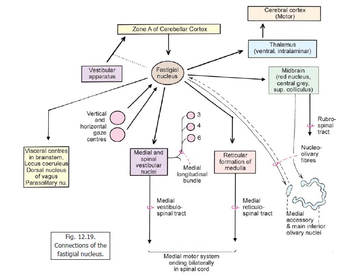
4. The fastigial nucleus also projects to the reticular formation of the medulla and influences motor neurons through the medial reticulospinal tract.The two tracts (medial vestibulospinal and medial reticulospinal) through which the vestibulocerebellum influences spinal motor neurons is called the ‘medial motor system’ (in distinction to the ‘lateral’ corticospinal system.
5. The fastigial nucleus sends efferents to the thalamus (ventral lateral and intralaminar nuclei). Fibres arising here reach the motor cortex and influence spinal motor neurons through corticospinal tracts.
B.Some fibres from the fastigial nucleus reach the midbrain. These fibres influence olivocerebellaroutput as follows.
a. Some fibres end in the red nucleus. From here fibres passing through the central tegmental tract reach the medial accessory olive.
b. Some fibres reach centres in the central grey matter (near the junction of the midbrain and the diencephalon). One of these is the nucleus of Darkschewitsch. Fibres arising in this region pass through the medial tegmental tract and reach the main inferior olivary nucleus. These inputs are excitatory for olivocerebellar impulses.
C.. The fastigial nucleus plays a role invisceromotor controlthrough connections with:
a) the parasolitary nucleus (lying near the solitary nucleus);
b) the locus coeruleus; and
c) the dorsal nucleus of the vagus nerve.
D. Control of eye movements
This topic has been discussed. Here we will consider the role of the cerebellum in control of finer eye movements (saccades and pursuit movements. The pathways involved are as follows.
1. The folium and tuber (of the cerebellar vermis), and also the flocculus, receive impulses of vision and those related to eye movements (through the inferior olivary complex). From here fibres project to the fastigial nucleus.
2. The fastigial nucleus also receives fibres from the vertical gaze centre (midbrain) and the horizontal gaze centre (pons).
3. We have seen that the fastigial nucleus projects to the medial and spinal vestibular nuclei. Fibres arising here pass through the medial longitudinal fasciculus to reach the oculomotor, trochlear and abducent nuclei. Fibres of the medial longitudinal fasciculus also reach the cervical spinal cord and influence muscles that rotate the neck (sternocleidomastoid, trapezius).
4. The fastigial nucleus is connected to the superior colliculus and can influence eye movements through it.
E. Control of olivocerebellar projection
The fastigial nucleus plays a role in controlling flow of impulses from the inferior olivary complex to the cerebellum. Some fibres from the fastigial nucleus project to the medial accessory olivary nucleus. These fibres are inhibitory.
Related Topics