Chapter: The Massage Connection ANATOMY AND PHYSIOLOGY : Lymphatic System
Components of the Lymphatic System
COMPONENTS OF THE LYMPHATIC SYSTEM
The lymphatic system is an anatomic system consist-ing of lymph vessels, lymph, specialized cells called lymphocytes (described in the section on Immunity), lymphoid organs, and collections of lymphoid tis-sue in different parts of the body.
Lymph Vessels
The lymphatic system is similar to the cardiovascular system because it, too, has vessels, often called lym-phatics. Lymphatics are present in almost all the re-gions of the body; however, they are absent from the central nervous system and such regions as the cornea, lens, cartilage, and epithelium that lack a blood supply.
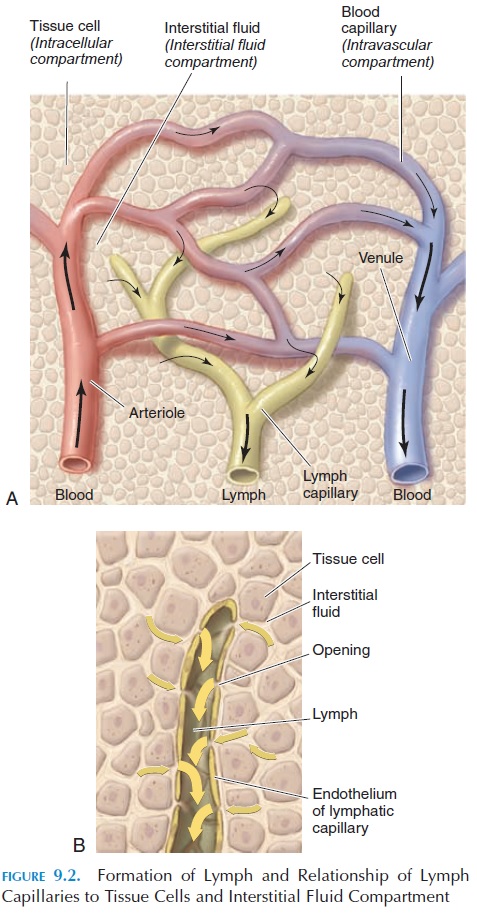
The smallest vessels—the lymphatic capillaries— arise as blind-ended tubes in the interstitial spaces (Figure 9.2). These capillaries have thinner walls, and they are larger than blood capillaries. The lymph cap-illaries are highly permeable and allow large particles to easily enter the vessel. The endothelial cells lining the capillaries have gaps that allow the particles to enter. In addition, the cells overlap with each other, with the overlap acting as one-way valves. Anchoringfilaments—proteins attached to the endothelialcells—also help adjust the width of the gaps. When there is more fluid inside the lymph capillaries, the width of the gap becomes smaller, allowing less fluid in and preventing backflow of fluid. When there is more fluid in the interstitial compartment, the an-choring filaments are pulled and the gap widens, al-lowing more fluid to enter the capillaries.
The lymph capillaries in the intestines (lacteals) are located in the center of the villi. The lymph in the lacteal carries a high fat content, giving the lymph a creamy, white appearance. Lymph flowing through the lacteals is referred to as chyle.

From the periphery, the networks of capillaries join and rejoin others to form larger lymphatic ves-sels (see Figure 9.3). The lymphatic vessels resembleveins, with an endothelium, smooth wall muscles, and adventitia. The inner lining of these large vessels is thrown into folds to form valves. Lymph vessels have numerous valves located every few millimeters, giving the vessels a beaded appearance. At various in-tervals, the lymph vessels open into lymphatic tissue called lymph nodes. Lymphatic vessels from the lymph nodes join others and progressively become larger until they communicate with two collecting vessels, the thoracic duct or left lymphatic duct, the largest of these vessels, and the right lymphaticduct. The lower end of the thoracic duct is enlargedand is known as the cisterna chyli.
The thoracic duct and the right lymphatic duct are located in the thoracic cavity. The thoracic duct is about 38–45 cm (15–17.7 in) long and runs parallel to the vertebral column. The right lymphatic duct is much shorter, about 1.5 cm (0.6 in) long. Both ducts open into the blood vessels in the neck (on the left and right side, respectively) at the junction of the subclavian and internal jugular vein. Thus, lymph is emptied into the blood circulation.
The thoracic duct collects lymph from the left side of the body and from the right side of the body infe-rior to the diaphragm. The right lymphatic duct col- lects lymph from the right side of the body superior to the diaphragm (i.e., the right side of the head and neck, the right upper limb, the right side of the tho-rax, the right lung, the right side of the heart, and part of the liver).
Lymph and Lymph Flow
The lymph or lymph fluid, a clear, pale yellow fluid, is the overflow fluid from the tissue spaces with the same composition as the interstitial fluid. It carries large proteins and waste from different parts of the body.
Lymph vessels, unlike blood vessels, do not have extensive smooth muscle around them to aid the flow.Also, the lymphatic system does not have a pump equivalent to the heart to circulate the fluid. This sys-tem, therefore, must rely on other mechanisms to move the fluid toward the neck. Lymph vessels have one-way valves to help direct the fluid. Lymph is also propelled to a large extent by the passive and active movements of skeletal muscles. In addition, the pul-sation of arteries lying close to the lymph vessels helps propel the lymph. Another important mecha-nism that draws the lymph upward is the respiratory movements. When a person inspires, the pressure drops in the thorax and increases in the abdomen. This difference in pressure is sufficient to “suck” the lymph into the thorax and the venous system. Changes in posture, passive compression, and mas-sage can also aid lymph flow. The smooth wall mus-cles of the lymphatic vessels also help move lymph by contracting when distended. The rate of flow in-creases with physical activity. It has been estimated that about 2–4 liters of lymphatic fluid and the equiv-alent of 25% to 50% of the total circulating plasma protein is returned to the circulation every day.
Lymph Nodes
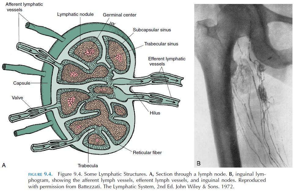
As lymph flows toward the blood circulation, at vari-ous points it passes through lymph nodes (see Figure 9.4). Lymph nodes are small organs of about 1–2 cm (0.4–0.8 in) that filter large particles and remove for-eign substances before lymph empties into the veins. They may be oval, round, elongated, or bean-shaped. Lymph nodes are also centers for proliferation of the lymphocytes. They are usually found in the subcuta-neous tissue (superficial nodes or muscle fascia and body cavities (deep nodes). Lymph nodes are numer-ous; there are more than 600 lymph nodes in the body.
Lymph nodes are often found in clusters, espe-cially in the axilla, groin, the side of the neck, thorax, and abdomen. The lymph nodes are located along the lymph vessels that lead from the tissue to the larger ducts. Each lymph node processes lymph from a spe-cific, adjacent anatomic site.
A lymph node is surrounded by a connective tissue capsule. The connective tissue of the capsule extendsinto the lymph node as trabeculae, dividing it into smaller compartments. The trabeculae and the mesh-work of connective tissue inside the lymph node pro-vide the framework for the lymph node and also help slow the flow of lymph as it passes through. The lymph node contains numerous lymphocytes and macrophages, types of white blood cells involved inthe defense of the body , arranged in clusters called germinal centers.
Tiny lymph vessels, called afferent vessels, bring the lymph into the lymph node. In the lymph node, the lymph flows through irregular channels (sinuses) that contain the white blood cells. Sinuses are present un-der the capsule (subcapsular sinuses), between the connective tissue (trabecular sinuses), and in the cen-ter of the lymph node (medullary sinuses). An efferent vessel takes lymphawayfrom the node after it isscreened by the cells located inside the node. These vessels emerge from the side of the lymph node through a small indentation known as the hilus.
If confronted by a foreign organism, white blood cells destroy the organism. At the same time, their multiplication is triggered. Certain lymphocytes pro-duce antibodies,proteins manufactured to destroy the specific organism (antigen). Antibody production causes lymph nodes that drain an infected area to be-come enlarged and painful during the infection, a condition called lymphadenitis. Sometimes, the lymph vessels are also inflamed and appear as thin, red streaks around the infected region, a condition called lymphangitis.
In addition to lymph vessels and lymph nodes, the lymphatic system includes the lymphoid organs; the thymus, spleen, and lymphoid tissue found in the ton-sils, appendix, and intestine.
The Thymus
The thymus (see Figure 9.5) is a flat, long structure with two lobes located in the mediastinum, inferior to the thyroid gland in the neck, posterior to the sternum. If you were able to pull your sternum forward and peek behind it, you would find it there! The thymus is surrounded by a connective tissue capsule. Similar to other lymphoid tissue, the thymus contains lympho-cytes, macrophages, and reticular epithelial cells.
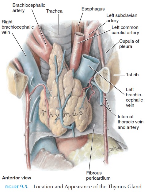
The thymus is fully developed at birth, and it con-tinues to grow until puberty. After puberty, it slowly decreases in size. The thymus is important in the de-velopment of the immune system. It is the first organ to begin manufacture of lymphocytes. The lympho-cytes processed in the thymus are called the T lym-phocytes. In the absence of the thymus, immunity issignificantly lowered. The thymus is considered an endocrine organ because it secretes the hormone thy-mosin.
The Spleen
The spleen (Figures 9.3 and 9.6) is an oval organ that is about as size of a clenched fist. It is located on the upper left quadrant of the abdomen, deep to ribs 9, 10, and 11, inferior to the diaphragm in contact with the stomach, splenic flexure of the colon, and the left kidney.
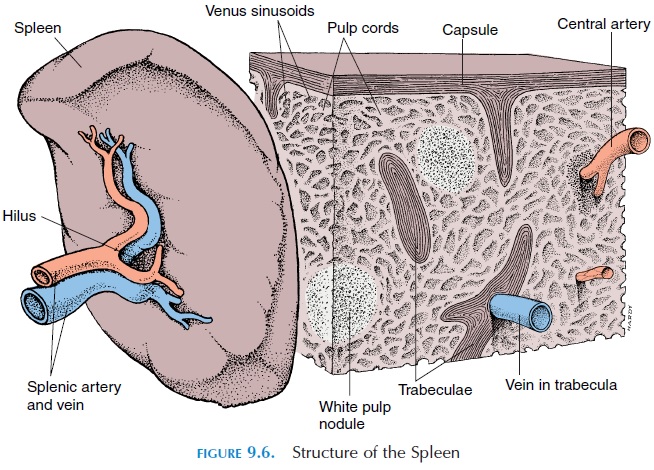
It is the largest mass of lymphoid tissue in the body. The spleen is covered by a connective tissue cap-sule from which trabeculae extend to the interior, pro-viding the framework. Microscopic examination re-veals that the spleen is composed of two, distinct kinds of tissue, the white pulp and red pulp. The white pulp contains an abundance of lymphocytes and macrophages, which destroy foreign tissue and manu-facture antibodies against them. These cells surround branches from the splenic artery. The red pulp consists of dilated veins called venous sinuses, which are filled and surrounded by cords of cells, consisting of red blood cells, macrophages, lymphocytes, plasma cells, and other white blood cells.
Tributaries of the splenic vein are closely associated with the red pulp. Within the red pulp, platelets are stored and injured and/or old red blood cells and platelets are destroyed. Before birth, the spleen also manufactures red blood cells.
The spleen is richly supplied with blood vessels; at any given time, it holds about 350 mL (11.8 oz) of blood. This volume of blood can be quickly sent back into the circulation when there is severe bleeding; therefore, the spleen serves as a blood reservoir. Un-fortunately, as a result of its vascularity, profuse bleeding can occur into the peritoneal cavity if the spleen is damaged as a result of trauma to the ab-domen. In such situations, the spleen is removed (splenectomy) to prevent the person from bleedingto death. Other structures, such as the bone marrow and liver, take over the functions of the spleen.
The Tonsils
The tonsils are collections of lymphoid tissue (lymphocytes and macrophages) located under the mucous membrane in the mouth and the back of the throat. They help protect against foreign agents that may enter the body through the nasal and oral cavities. The tissues located on either side of the throat are known as the palatine tonsils.Those located in the throat near the posterior opening of the nasal cavity are the pharyngeal tonsils, or ade-noids. The paired collections of lymphoid tissue at thebase of the tongue are known as lingual tonsils.
Intestinal Lymphoid Tissue
Lymphoid tissue located in the submucosa of the in-testines and in the appendix defends the body against foreign agents that may enter the system through the gut. In the gut, the lymphoid tissues are scattered in patches. In the intestine, these patches are known asPeyer’s patches.
Related Topics