Chapter: Medical Physiology: The Eye: II. Receptor and Neural Function of the Retina
Color Vision: Tricolor Mechanism of Color Detection, Color Blindness
Color Vision
From the preceding sections, we have learned that different cones are sensitive to different colors of light. This section is a discussion of the mechanisms by which the retina detects the different gradations of color in the visual spectrum.
Tricolor Mechanism of Color Detection
All theories of color vision are based on the well-known observation that the human eye can detect almost all gradations of colors when only red, green, and blue monochromatic lights are appropriately mixed in different combinations.
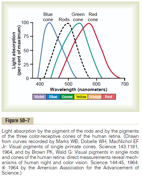
Spectral Sensitivities of the Three Types of Cones. On thebasis of color vision tests, the spectral sensitivities of the three types of cones in humans have proved to be essentially the same as the light absorption curves for the three types of pigment found in the cones. These curves are shown in Figure 50–7 and slightly differ-ently in Figure 50–9. They can explain most of the phe-nomena of color vision.
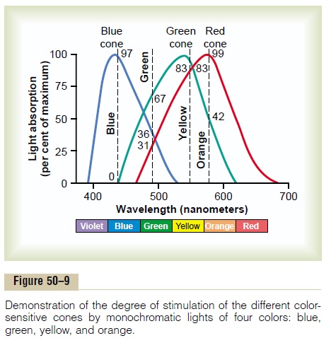
Interpretation of Color in the Nervous System. Refer- ring to Figure 50–9, one can see that an orange monochromatic light with a wavelength of 580 nanometers stimulates the red cones to a stimulus value of about 99 (99 per cent of the peak stimulation at optimum wavelength); it stimulates the green cones to a stimulus value of about 42, but the blue cones not at all. Thus, the ratios of stimulation of the three types of cones in this instance are 99:42:0. The nervous system interprets this set of ratios as the sensation of orange. Conversely, a monochromatic blue light with a wavelength of 450 nanometers stimulates the red cones to a stimulus value of 0, the green cones to a value of 0, and the blue cones to a value of 97. This set of ratios—0:0:97—is interpreted by the nervous system as blue. Likewise, ratios of 83:83:0 are inter-preted as yellow, and 31:67:36 as green.
Perception of White Light. About equal stimulation ofall the red, green, and blue cones gives one the sensa-tion of seeing white. Yet there is no single wavelength of light corresponding to white; instead, white is a com-bination of all the wavelengths of the spectrum. Fur-thermore, the perception of white can be achieved by stimulating the retina with a proper combination of only three chosen colors that stimulate the respective types of cones about equally.
Color Blindness
Red-Green Color Blindness. When a single group of color-receptive cones is missing from the eye, the person is unable to distinguish some colors from others. For instance, one can see in Figure 50–9 that green, yellow, orange, and red colors, which are the colors between the wavelengths of 525 and 675 nanometers, are normally distinguished from one another by the red and green cones. If either of these two cones is missing, the person cannot use this mechanism for distinguishing these four colors; the person is especially unable to distinguish red from green and is therefore said to have red-green colorblindness.
A person with loss of red cones is called a protanope; the overall visual spectrum is noticeably shortened at the long wavelength end because of a lack of the red cones. A color-blind person who lacks green cones is called a deuteranope; this person has a perfectly normal visual spectral width because red cones are available to detect the long wavelength red color.
Red-green color blindness is a genetic disorder that occurs almost exclusively in males. That is, genes in the female X chromosome code for the respective cones. Yet color blindness almost never occurs in females because at least one of the two X chromosomes almost always has a normal gene for each type of cone. Because the male has only one X chromosome, a missing gene can lead to color blindness.
Because the X chromosome in the male is always inherited from the mother, never from the father, color blindness is passed from mother to son, and the mother is said to be a color blindness carrier; this is true in about 8 per cent of all women.
Blue Weakness. Only rarely are blue cones missing,although sometimes they are underrepresented, which is a genetically inherited state giving rise to the phe-nomenon called blue weakness.
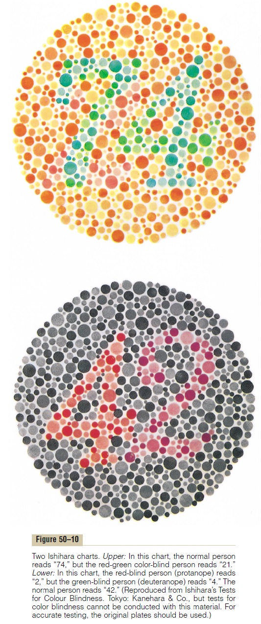
Color Test Charts. A rapid method for determining colorblindness is based on the use of spot charts such as those shown in Figure 50–10. These charts are arranged with a confusion of spots of several different colors. In the top chart, the person with normal color vision reads “74,” whereas the red-green color-blind person reads “21.” In the bottom chart, the person with normal color vision reads “42,” whereas the red-blind person reads “2,” and the green-blind person reads “4.”
If one studies these charts while at the same time observing the spectral sensitivity curves of the different cones depicted in Figure 50–9, it can be readily under-stood how excessive emphasis can be placed on spots of certain colors by color-blind people.
Related Topics