Chapter: Human Nervous System and Sensory Organs : Brain's Cerebrovascular and Ventricular Systems
Choroid Plexus - Cerebrospinal Fluid Spaces

Choroid Plexus
Lateral Ventricles (A, C)
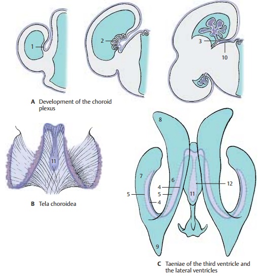
The choroid plexus consists of convolutions of blood vessels protruding into the ventricles from specific parts of the wall. The area of the wall adhering to the medial sur-face of the hemisphere (choroid lamina) (A1) becomes thinner during embryonic development and is pushed into the ventricular lumen by the vascular loops of the pia mater (A2). At the beginning of development, all vascular convolutions are covered by a thin layer of hemispheric wall. The latter finally turns into a layer of epithelial cells, the plexus epithelium. Thus, the adult choroid plexus consists of two components, namely, the vascularized con-nective tissue of the pia mater and the plexus epithelium (transformed hemi-spheric wall). Once the choroid plexus has invaginated into the ventricular cavity, it re-mains connected with the external pia mater only through a narrow channel, the choroid fissure(A3). When the choroidplexus is removed, the thinned parts of the hemispheric wall tear away at this location. The line of attachment is called taenia(band). One such line is attached to the for-nix and to the fimbria of the hip-pocampus; it is known as the taenia fornicis (taenia of the fornix) (C4).The other line runs along the lamina affixa and is known as the taeniachoroidea (choroid line) (C5).
Owing to the rotation of the hemispheres, the choroid plexus forms a semi-circle along the medial wall of the ventricle, reaching from the interventricular foramen (foramen of Monro) via the central part (C6) to the inferior horn (C7). The anterior horn (C8) and the posterior horn (C9) do not con-tain a plexus.
Tela choroidea (A – C)
When the hemispheres overgrow the dien-cephalon, the leptomeninges of both parts of the brain come to lie on top of each other to form aduplication(A10), the tela choroidea (B), a connective-tissue plate spanning between the telencephalic hemispheres and the diencephalon. At its lateral margins, the pia mater forms the vascular convolution for the choroid plexus of the lateral ven-tricles. In the middle, the tela covers the roof of the third ventricle (tela choroidea of thethird ventricle) (BC11). In this region, tworows of convoluted blood vessels protrude into the lumen of the third ventricle and form the choroid plexus of the third ventricle. When removing the ventricular roof, the taenia thalami (C12) remains as line of at-tachment running along the medullary stria across the thalamus.
Fourth Ventricle (D, E)
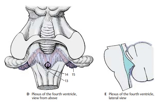
Above the fourth ventricle, the tela choroideaof the fourth ventricle is also formed as adu-plication of the pia mater by apposition ofthe lower surface of the cerebellum to the surface of the rhombencephalon (E). The roof of the rhombencephalon becomes re-duced to a thin epithelial layer and is pushed into the ventricle by the vascular loops originating from the tela. The tela choroidea of the fourth ventricle consist of pia mater only, as the arachnoidea does not adhere to the surface of the cerebellum but spans the cerebellomedullary cistern. At the obex (D13), the attachment of the tela abovea narrow medullary fold, lies the medianaperture (foramen of Magendie) (D14). Onboth sides open the lateral apertures (foramina of Luschka), through which the lateral end of the choroid plexus protrudes (Bochdalek’s flower basket) (D15).
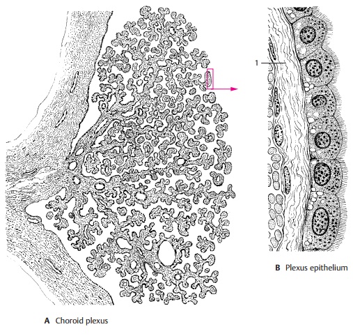
The treelike ramification of the choroid plexus (A) creates a very large surface. Each branch contains one or more vessels, such as arteries, capillaries, and thin-walled venous caverns. The vessels are surrounded by a loose meshwork of collagen fibers (B1) which, in turn, is covered by the plexus epithelium (B). The plexus epithelium con-sists of a single layer of cuboidal cells that carry a fine brush border on their apical sur-face. Their cytoplasm contains vacuoles and coarse granules as well as inclusions of lipid and glycogen.
The choroid plexus is the site of cerebrospi-nal fluid production. Fluid is transferredfrom the vascular system of the choroid plexus through the epithelium into the ven-tricles. Whether this occurs through secre-tion by the plexus epithelium or through dialysis (a form of filtration) has yet to be established.
Like the pia-arachnoid and dura, the choroid plexus is richly innervated (the meninges are innervated by the trigeminal nerve, vagus nerve, and autonomic fibers). The choroid plexus and meninges are therefore sensitive to pain, whereas the brain tissue itself is largely insensitive.
Related Topics