Chapter: Case Study in Obstetrics and Gynaecology: General Gynaecology
Case Study Reports: Abnormal Cervical Smear
ABNORMAL CERVICAL SMEAR
History
A
28-year-old woman attends
the colposcopy clinic
after an abnormal smear test. She
is very anxious as she thinks
that she might
have cervical cancer.
The smear is reported as ‘severe dyskaryosis’. She
had a previous normal result
at age 25 years. She
has not had
any postcoital or intermenstrual bleeding.
Her
first sexual relationship started at the age of 14 years
and she has had several
part- ners since then.
She lives with her current
partner who she has been with for 3 years.
She was diagnosed with genital herpes
several years ago but has not had any attacks
for at least 3 years. She
smokes 15–20 cigarettes per day and
drinks only at the weekends.
She has an intrauterine
contraceptive device in situ.
Examination
The
cervix is macroscopically normal. At colposcopy, acetic acid is applied and
an irregu- lar white
area is apparent to the left
of the os.
Lugol’s iodine is applied and
the same area stains pale while the
rest of the
cervix stains dark
brown. A biopsy
is taken
Questions
·
How should this patient be managed?
Answer
The
colposcopy findings show an abnormal
area on the left of the cervix.
The abnormal tissue stains
white with acetic
acid because abnormal
cells have high-density nuclei which take up the acetic
acid more than normal cells.
In contrast, abnormal
cells have lower glycogen content than normal
cells and stain
less well, remaining pale when iodine
is applied.
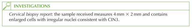
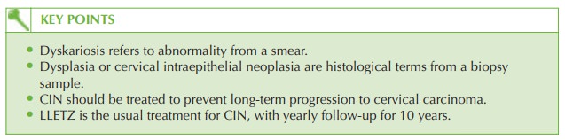
The
diagnosis is of CIN3 (cervical intraepithelial neoplasia). This is a tissue diagnosis as opposed to dyskaryosis which is an observation of cells from
a smear. The
degree of dyskaryosis and
CIN often correlate, but a dyskaryosis report is not
a diagnosis.
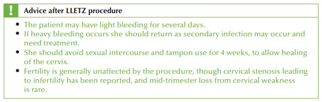
Management
CIN
needs to be treated to prevent progression over several years
to cervical carcinoma. The commonest treatment is
large-loop excision of the transformation zone (LLETZ) – removal of abnormal cervical tissue with a diathermy loop.
Most women tolerate this under local anaesthetic. The LLETZ sample needs to be examined
histologically both to confirm removal of all
the abnormal tissue,
and to ensure
that there is not a focus of car-
cinoma within the sample.
Follow-up smear should be performed in 6 months,
and thereafter yearly
smears for 10 years.
Related Topics