Chapter: Microbiology and Immunology: Morphology and Physiology of Bacteria
Capsule and Slime Layer - Structure and Functions of Bacterial Cell Envelope
Structure and Functions of Bacterial Cell Envelope
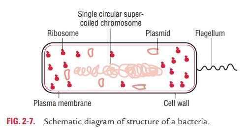
The outer layer or cell envelope provides a structural and physiological barrier between the protoplasm (inside) of the cell and the external environment. The cell envelope protects bacteria against osmotic lysis and gives bacteria rigidity and shape. The cell envelope primarily consists of two components: a cell wall and cytoplasmic or plasma membrane. It encloses the proto-plasm, which consists of (i) cytoplasm, (ii) cytoplasmic inclu-sions (mesosomes, ribosomes, inclusion granules, vacuoles), and (iii) a single circular DNA (Fig. 2-7).
Capsule and Slime Layer
Many bacteria, both Gram-positive and Gram-negative, possess a gel-like layer outside the envelope when growing in their natural environments. When a gel-like layer forms a well-defined condensed layer around the bacterial envelope, it is called a capsule and is demonstrable by a light microscope. When this gel-like layer is narrower, detectable only by indirect serological methods or by electron microscope but not by light microscope, it is called a microcapsule. An amorphous viscid colloidal material secreted by some bacteria extracellularly is termed as loose or free slime or glycocalyx.
◗ Capsule
The capsule is mostly made up of polysaccharides, often referred to collectively as exopolysaccharides. Exopolysaccharides are some-times neutral homopolysaccharides (e.g., the glucans and fruc-tans of many oral streptococci) or negatively charged (Table 2-4).
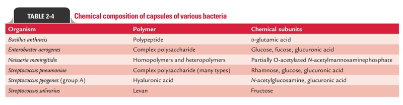
However, Bacillus anthracis has a capsule comprising of polyamino acids, such as D-glutamic acid. The D-glutamic acid is probably analogous to the negatively charged polysaccharide capsule.
Demonstration of capsule
The capsule is fully hydrated and can be demonstrated by light microscopy in either living or stained bacteria as follows:
Special capsular staining methods: These include Welch methodand M’Faydean capsule stain. Welch method uses copper as mordant. This involves treatment of fixed smear with hot crystal violet solution followed by rinsing with copper sulfate solution. The latter is used to remove excess stain because conventional washing with water would dissolve the capsule. The copper salt also gives color to the background, with the result that the cell and background appear dark blue and the capsule of the bacteria a much paler blue. M’Fadyean capsulestain, using polychrome methylene blue stain, is a frequentlyused method for demonstration of capsule ofB. anthracis.
Negative staining with India ink: Also known as wet India inkmethod. It is the simplest way to demonstrate capsule. It is carried out by mixing a suspension of bacteria with an equal volume of Indian ink on a slide, covering with a cover slip, and then examining it under microscope. The capsule appears as a clear zone around the cell. This method is useful for improving visualization of encapsulated bacteria in clinical samples, such as blood or cerebrospinal fluid.
Serologic methods: Since capsules are antigenic, they can bedemonstrated by serologic methods. Quellung’s reaction is such a serological method for demonstration of capsule. When a suspension of bacterium is mixed with its specific anticapsular serum and methylene blue and examined under microscope, the capsule becomes very prominent and appears swollen due to increase in refractoriness. This method is useful for rapid identification of capsular serotypes of S. pneumoniae, N.meningitidis, H. influenzae,Yersinia, Bacillus, etc.
◗ Slime layer
Slime layer (S-layer) is a structured paracrystalline protein layer shown by electron microscopy. These are generally composed of a single kind of protein molecule, sometimes with carbohy-drates attached. They are resistant to proteolytic enzymes and protein-denaturing agents. The slime layer protein protects the cell from wall-degrading enzymes and bacteriophages. It plays an important role in the maintenance of cell shape, and it may be involved in cell adhesion to host epidermal surfaces.
Related Topics