Chapter: Psychology: The Brain and the Nervous System
Building Blocks of the Nervous System
BUILDING BLOCKS OF THE
NERVOUS SYSTEM
Descartes’ views were based
largely on conjecture, because in his time scientists knew little about how the
nervous system functions. Today, roughly five centuries later, we know far
more and our
theorizing can be
correspondingly more sophisticated. In fact, the data and research
tools now available have allowed the development of a new and rapidly growing
field called neuroscience—a multidisciplinary
effort that seeks to understand
the nature, function,
and origins of
the nervous system. Neuroscience draws some of its
insights from psychology but also draws on biol-ogy, computer
science, medicine, and
other fields. The subject matter
of neuro-science is
similarly broad, ranging
from fine-grained research
projects that scrutinize molecular
interactions inside individual
nerve cells to
much broader efforts asking how
large tracts of neural tissue give rise to conscious experience.
Let’s be clear from the start
that studying the brain is, to say the least, an enormously daunting task.
Within the human brain, the total number of neurons—the individual cells that act as the main information
processors of the nervous system—has been esti-mated to be as high as 100
billion, roughly the number of stars in the Milky Way, and each of these
neurons connects to as many as 50 thousand others (Nauta & Feirtag, 1986).
The brain also contains another type of cell, glia, whose function we’re just
begin-ning to understand. In some parts of the brain, glia outnumber the
neurons by 10 to 1. All these cells, and all their interconnections, are
contained within an organ that weighs only 3 to 4 pounds—leading many writers to
suggest that the human brain is the most complex object in the universe.
As a first step in understanding
this complexity, let’s look at the basic building blocks of the nervous
system—the neurons and glia. We’ll then turn to the nerveimpulse—the means through which individual neurons communicate
with each other.
The Neuron
The neuron is a cell that
specializes in sending and receiving information. Neurons typically have three
main parts: the dendrites, the cell body (or soma), and the axon
(Figure 3.3). The dendrites, the “input” side of the neuron, receive signals
from many other neurons. In most neurons, the dendrites are heavily branched,
like a thick and tan-
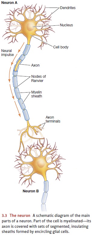
gled bush. The cell body contains
the neuron’s nucleus and all the elements needed for the normal metabolic
activities of these cells. The axon, finally, is the “output” side of the
neuron and sends neural impulses to other neurons. The axon usually extends
outward from the cell body like a wispy thread, and it may fork into several
branches at its end.
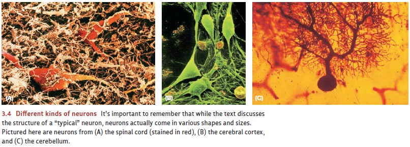
Neurons come in many shapes and
sizes (Figure 3.4). Their cell bodies vary from 5 to about 100 microns in
diameter (1 micron = 1/1,000 millimeter). In comparison, the aver-age human
hair has a diameter of about 100 microns. Neurons’ dendrites are typically
short—say, a few hundred microns. Axons, however, can be much longer. For
example, the longest axons in humans are those of the motor neurons, which transmit neural impulses from the brain to the
muscles. The motor neurons that allow us to wiggle our toes are, in most
people, more than a meter long. As a result, these particular neurons, with
their small cell bodies connected to very long axons, have roughly the same
proportions as a basket-ball attached to a garden hose a mile and a half long.
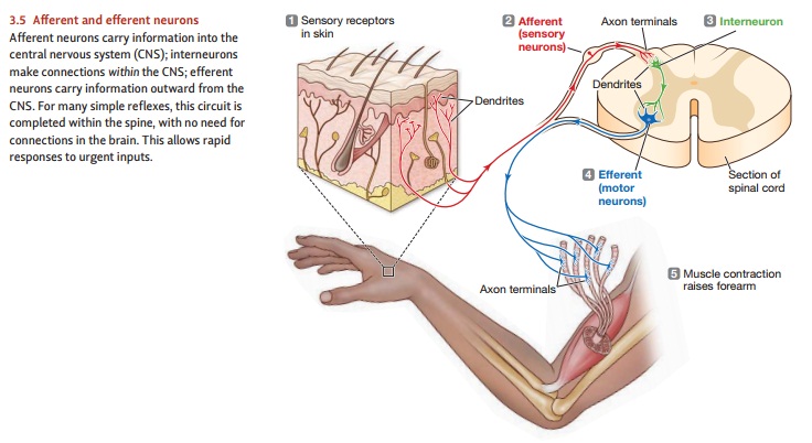
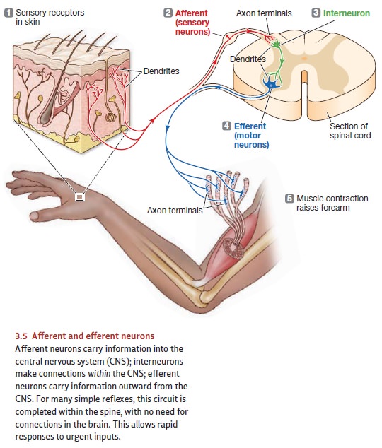
The motor neuron carries an efferent signal 1that allows the brain
to control the mus-cles. Efferent neurons in general carry information from the
brain to some destination outside the brain. Other neurons convey information
inward; these afferent neurons keep
the nervous system informed about both the external world and the body’s
internal environment (Figure 3.5).* Some of the afferent neurons are attached
to spe-cialized receptor cells that respond to external energies such as
pressure, chemical changes, light, and so on. These receptor cells translate
(more technically, transduce) the
physical stimuli into electrical changes, which then trigger a nervous impulse
in other neurons.
It turns out, though, that
roughly 99% of the brain’s nerve cells are neither afferent nor efferent
neurons. Instead, they’re neurons that make connections within the central nervous system, and they’re divided into two
types: The projection neurons link
one area of the central nervous system to some other (perhaps distant) area; to
perform this function, these nerve cells typically have long axons. The interneurons, in contrast, make “local”
connections within the nervous system and usually have either very short axons
or none at all. (Be aware that some neuroscientists use the term interneuron to refer to both of these
nerve cell types—e.g., Kolb & Whishaw, 2009.)
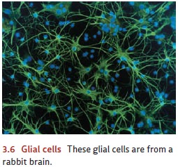
Glia
What about the other type of
cells that make up the brain—the glia
(Figure 3.6)? For many years, scholars thought the glia played only a few
roles—holding the neurons in place and supplying them with nutrients and
oxygen. Indeed, this second-class status is reflected in the word glia itself, which comes from the Greek
for “glue” or “slime.”
It turns out, however, that glia
have a rather broad set of crucial functions. First, as we’ve long supposed,
glia do provide the nourishment for the neurons—but we’ve also learned that
glia play a key role in controlling
the nutrient supply. For example, the sugar glucose
is the main fuel for the nervous system, but most of the energy neurons need
does not come from glucose directly. Instead the glia convert the glucose into
another molecule, called lactate, that feeds the neurons. The glia are also
sensitive to the activity level in each neuron and increase the blood flow
(providing more oxygen and fuel) whenever the neurons in the brain region
become more active (Rouach, Koulakoff, Abudara, Willecke, & Giaume, 2008).
The glia also play a central role
in the brain’s development. Before
birth and in the months afterward, the human brain grows at a remarkable rate
as its cells rapidly reproduce and differentiate. The newly created neurons
then migrate from one posi-tion in the brain to another, moving at a speed of
up to 1 millimeter each day. This migration is guided by glia, acting as
guidewires—much like beanpoles guiding the growth of bean shoots in the garden.
Then, once the neurons have reached their des-tinations and established the
appropriate connections, the glia produce chemicals that help to shut down the
process of neural growth. In this way, the glia ensure a rel-atively stable
pattern of connections.
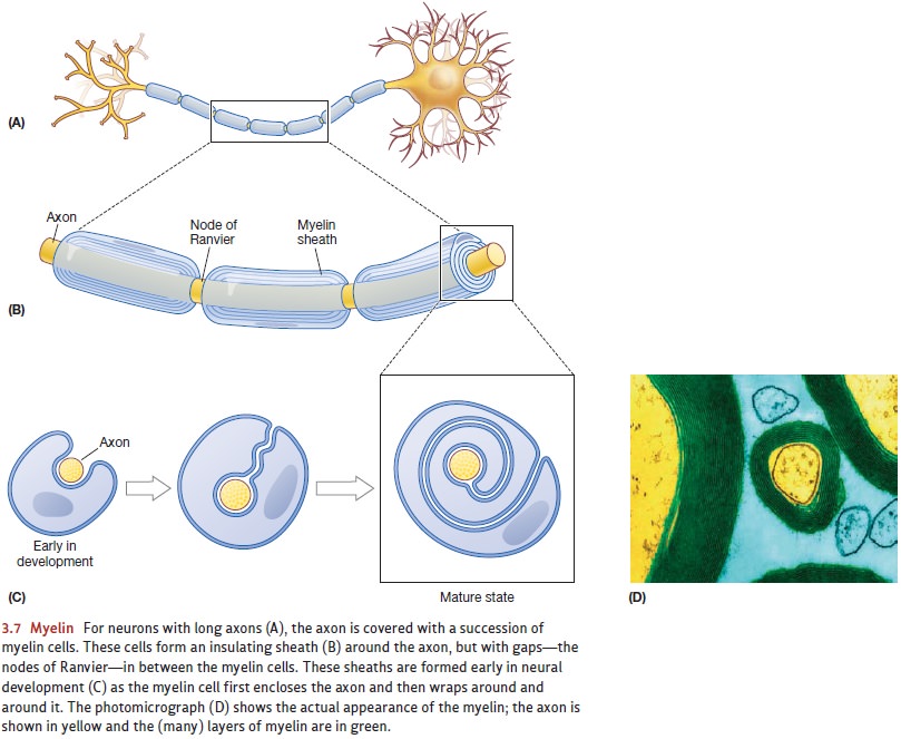
Yet another function of glial
cells is to increase the speed of neuronal communi-cation. The glia that
accomplish this are mostly made of a fatty substance known as myelin; and soon after birth, these
glia start to wrap themselves around the axons ofneurons—especially the longer
axons that span greater distances and thus need greater transmission speed
(Figure 3.7). Each of the “wrappers” in this myelin sheath covers a portion of the axon, and soon the entire
length of the axon is covered by a series of these wrappers. Crucially, though,
there are gaps—called the nodes ofRanvier—between
the successive wrappers, and it’s this combination of wrappersand gaps that
speeds up the nerve impulses traveling along these myelinated axons. (We’ll discuss later why the myelination speeds up the impulse.)
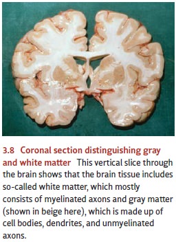
Myelin is white—which explains
why, when you look at a brain, you’ll see a mix of white matter and gray matter
(Figure 3.8). The white matter
consists of the myelinated axons traversing long distances either within the
brain or to and from the body. Conversely, gray
matter consists of cell bodies, dendrites, and the unmyelinated axons.
The glia may also have other jobs
to do. For example, several recent studies suggest that glia can “talk back” to
the neurons, sending signals that help regulate the strength of connections
between adjacent neurons. In some circumstances, glia can also release
chemicals that increase the reactivity of neurons. This is usually helpful—it
makes the nervous system more sensitive to important inputs—but, unfortunately,
this same mechanism can sometimes make the neurons too reactive. In the
extreme, this increased reactivity may be the source of so-called neuropathic
pain—a condition in which people suffer extreme pain in response to even a mild
touch—and it may also play a role in the development of epilepsy and several
other illnesses (G. Miller, 2005a).
Still other evidence suggests
that the glial cells themselves may constitute a separate, slow signaling
system within the brain. Glial cells are known to respond to various
elec-trical, chemical, and mechanical stimuli. They also form networks that
communicate with each other and that may modulate the activity level of neurons
nearby. The extent to which these networks of glia interact with neurons in the
brain has not yet been determined, but it may be considerable (Bullock et al.,
2005; Gallo & Chitajallu, 2001; Newman & Zahs, 1998; Verkhratsky,
1998).
Related Topics