Characteristic features, Classification, Economic importance, structure, Reproduction - Bryophytes | 11th Botany : Chapter 2 : Plant Kingdom
Chapter: 11th Botany : Chapter 2 : Plant Kingdom
Bryophytes
Bryophytes
Amphibians of Plant Kingdom
In the previous chapter we noticed a wide range of thallus organization in Algae. Majority of them are aquatic.
The development of heterotrichous habit,
development of parenchyma tissue, dichotomous branching in some algae supports
the view that colonization of plants in land occurred in the past. Bryophytes
are simplest and most primitive plant groups descended from alga – like
ancestors. They are simple embryophytes. Let us learn about the structure and
reproduction of these primitive land plants called Bryophytes in detail.
Bryophytes are simplest land inhabiting cryptogams
and are restricted to moist, shady habitats. They lack vascular tissue and
hence called ‘Non- vascular cryptogams’.
They are also called as ‘amphibians of
plant kingdom’ because they need
water for completing their life
cycle.
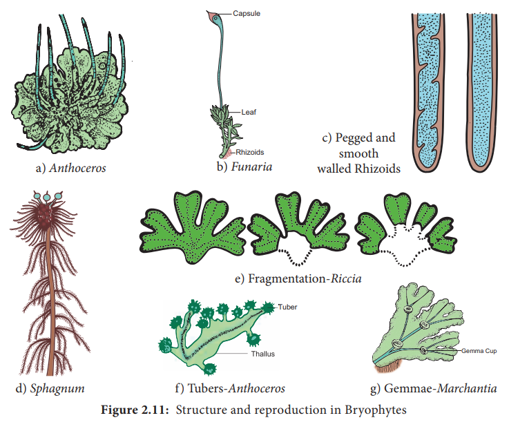
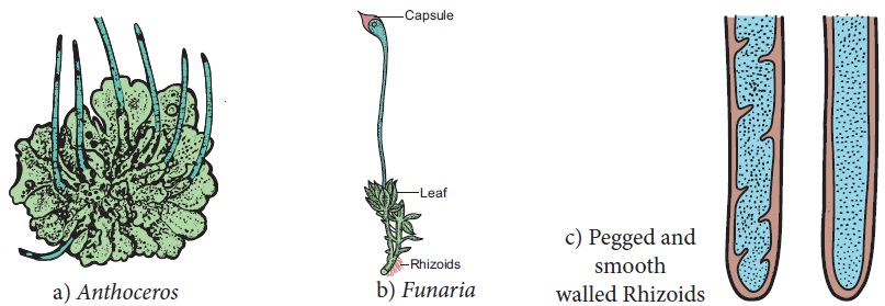
1. General characteristic features
•
The plant body of bryophyte is gametophyte and is
not differentiated into root, stem and leaf like structure.
•
Most of them are primitive land dwellers. Some of
them are aquatic (Riella, Ricciocarpus).
•
The gametophyte is conspicuous, long lived phase of
the life cycle. Thalloid forms are present in liverworts and Hornworts. In
Mosses leaf like, stem like structures are present. In Liverworts thallus grows
prostrate on the ground and is attached to the substratum by means of rhizoids.
Two types of rhizoids are present namely smooth walled and pegged.
Multicellular scales are also present. In Moss the plant body is erect with
central axis bearing leaf like expansions. Multicellular rhizoids are present.
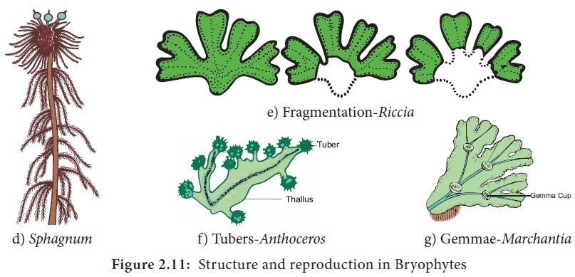
The structure and reproduction in Bryophytes is given in Figure 2.11
•
Vascular tissue like xylem and phloem are
completely absent, hence called ‘Non vascular cryptogams’.
•
Vegetative reproduction takes place by the
formation of adventitious buds (Riccia
fluitans) tubers develop in Anthoceros.
In some forms small detachable branches or brood bodies are formed, they help
in vegetative reproduction as in Bryopteris
fruticulosa. In Marchantia propagative organs
called gemmae are formed and help in reproduction.
• Sexual
reproduction is Oogamous. Antheridia and Archegonia are produced in a
protective covering and are multicellular
• The
antheridia produces biflagellate antherozoids which swims in thin film of water
and reach the archegonium and fuse with the egg to form diploid zygote.
•
Water is essential for fertilization.
• The zygote is the first cell of the sporophyte generation. It undergoes mitotic division to form multicellular undifferentiated embryo. The embryogeny is exoscopic (the first division of the zygote is transverse and the apex of the embryo develops from the outer cell) . The embryo divides and give rise to sporophyte.
• The sporophyte is dependent on gametophyte.
•
It is differentiated into three recognizable parts
namely foot, seta and capsule. Foot is the basal portion and is embedded in the
gametophyte through which water and nutrients are supplied for the sporophyte.
The diploid spore mother cells found in the capsule region undergoes meiotic
division and give rise to haploid spores. Bryophytes are homosporous. In some
sporophytes elaters are present and help in dispersal of spores (Example: Marchantia). The spores germinate to produce gametophyte.
•
The zygote, embryo and the sporogonium constitute
sporophytic phase. The green long living haploid phase is called gametophytic
phase The haploid gametophytic phase alternates with diploid sporophyte and
shows heterologous alternation of generation.
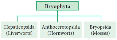
2. Classification of Bryophytes
Proskauer in the year 1957 classified Bryophytes
into 3 Classes namely
i.
Hepaticopsida
(Riccia, Marchantia, Porella and Riella) ii. Anthocerotopsida (Anthoceros
and Dendroceros) iii. Bryopsida (Funaria, Polytrichum and Sphagnum).
The outline of the classification is given below
Class: Hepaticopsida
They are lower forms of Bryophytes. They are more
simple in structure than mosses and more confined to damp and shady places.
They have an undifferentiated thallus. Protonernal stage is absent. Sporophyte
is very simple and short lived. In some the foot and seta are absent. Example Riccia.
Class: Anthocerotopsida
Gametophyte is undifferentiated thallus. Rhizoids
are unicellular and unbranched. Protonemal stage is absent. Sporophyte is
differentiated into foot and capsule and seta is absent Example: Anthoceros.
Class: Bryopsida
These are higher forms in which the gametophyte is differentiated into ‘stem’ like and’leaf’ like parts and the former showing radial symmetry. Rhizoids are multi-cellular and branched. Protonemal stage is present. Sporophyte is differentiated into foot, seta and capsule. They have a more differentiated structure than liverworts. They often form dense cushions. Example: Funaria.
3. Economic importance
A large amount of dead thallus of Sphagnum gets accumulated and compressed, hardened to form peat. In
northern Europe peat is used as fuel in commercial scale (Netherlands) . Apart
from this Nitrates, brown dye and tanning materials are derived from peat. Sphagnum and peat are also used in
horticulture as packing material because of their water holding capacity. Marchantia polymorpha is used to cure
pulmonary tuberculosis. Sphagnum, Bryum and Polytrichum are used as
food. Bryophytes play a major role in soil formation through succession and
help in soil conservation.
4. Marchantia
Class - Hepaticopsida
Order – Marchantiales
Family - Marchantiaceae
Genus - Marchantia
Marchantia
grows in
cool moist shady places. Marchantia polymorpha is the common
species.
Gametophyte
The plant body of Marchantia is a gametophyte. It is prostrate,dorsiventral and
dichotomously branched. The thallus on the dorsal surface possess conspicuous
median midrib which is marked by a shallow groove on dorsal surface. The dorsal
surface appears to have rhomboidal or polygonal diamond shaped areas which
indicate the outline of the underlying air chambers of the thallus (Figure 2.12).
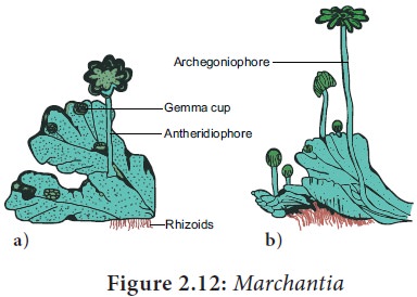
Figure
2.12: Marchantia
a)
Thallus with antheridiophore
b)
Thallus with archegoniophore
Internal structure of Thallus
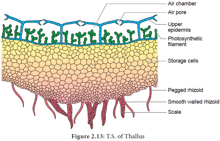
The epidermis has the upper and lower layers. The upper epidermis is single layered with thin walled parenchymatous cells. The cells possess chloroplasts. The upper epidermis is interrupted by many barrel shaped air pores which communicate with the air chambers. The pore is surrounded by 4 to 8 superimposed tiers of cells. Below the upper epidermis a number of air chambers are present in a single horizontal layer.The air chambers are separated from one another by partitions which extend from the epidermis to the floor of the air chambers. The floor of the chambers bears simple or branched green filaments. The cells of the filaments are involved in photosynthesis. The photosynthetic region is followed by storage region. It is made up of several parenchymatous cells arranged without intercellular spaces. The cells of this region contain starch grains and protein granules. The lower epidermis possesses rhizoids and multicellular scales.
Reproduction
Marchantia
reproduces
by vegetative and sexual methods.
1.Vegetative Reproduction takes place by
progressive death and decay of thallus, formation of adventitious branches and
by germination of gemmae. Death and decay of the thallus starts from posterior
end .When it reach the point of dichotomy , two apical parts of the thallus get
separated. Each one develops into an independent thallus. Adventitious branches
are produced on the ventral surface of the gametophyte. The branches get
separated from the parent thallus and grow into independent gametophytes.
Gemmae are specialized multicellular asexual reproductive bodies. They are
formed in small cupules known as gemma cups, present on the dorsal surface of the
thallus. Usually the gemmae present on the male thallus form male plants and
those on the female thallus give rise to female plants (Figure 2.14).
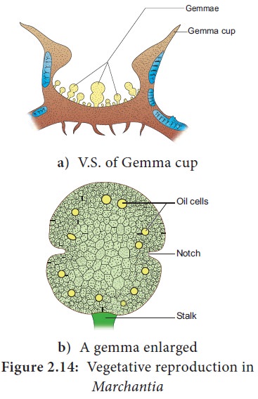
Vegetative reproduction in Marchantia
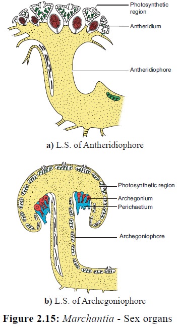
1. Sexual reproduction:
In Marchantia,
sex organs are borne on special stalked receptacles called the gametophores.
Those bearing antheridia are called antheridiophores and archegonia bearing
structures are called archegoniophores (Figure
2.15). Marchantia is heterothallic or
dioecious. i.e., male and female
receptacles are present on different thalli. The sex organs in bryophytes are
multicellular. The male sex organ is called antheridium. It produce
biflagellate antherozoids. The female sex organ is flask shaped called
archegonium and produces a single egg. Water is essential for fertilization.
The antherozoids are released into water and are attracted towards archegonium
through chemotaxis. Although many antherozoids enter the archegonium, only one
fuses with the egg to form zygote. The zygote represent the first cell of the
sporophytic generation. Zygote develops in to a multicellular structure called
sporophyte. (Figure 2.16).
The sporophyte is not free-living but attached to
the photosynthetic gametophyte and derives nutrition from it. Sporophyte is
differentiated into foot, seta and capsule. The foot is bulbous and is embedded
in the gametophyte. It derives nutrition from the gametophyte and transfers to
the sporophyte. Seta is short and connects foot and capsule. The capsule
consists of single layered jacket layer and encloses numerous haploid spores
and elaters. The capsule is covered by protective covering called calyptra. On
maturation the capsule dehisces and spores are released. Elaters helps in the
dispersal of spores. The spores under favourable conditions germinate and
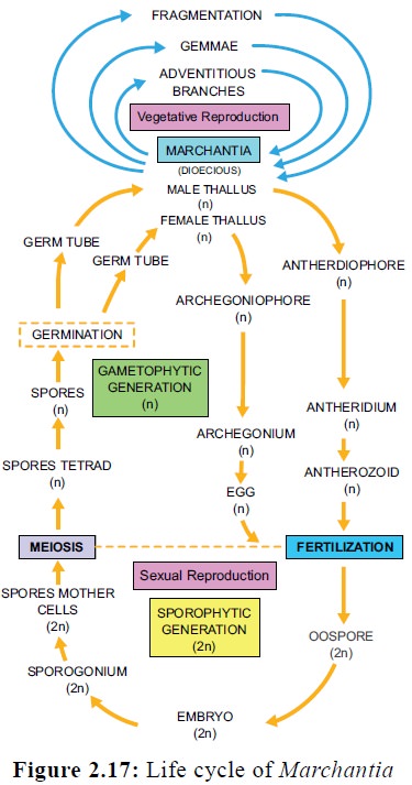
develop into new gametophyte. The haploid gametophytic phase alternates with
diploid sporophytic phase, thus the life cycle of Marchantia shows alternation of generation (Figure 2.17).
5. Funaria
Class – Bryopsida
Order- Funariales
Family – Funariaceae
Genus – Funaria
Funaria is commonly called ‘cord moss’. It is distributed throughout the world. Funaria hygrometrica is the common species. It grows in close tufts on rocks, trunks of trees, damp walls and damp soils. They help in the process of soil formation (Pedogenesis).
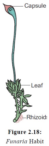
External features
The plant body is a gametophyte. It is small, 1 to
3 cm high and consists of slender erect radial stem covered with small, simple
leaf like structures arranged in a spiral manner. The gametophyte is attached
to the substratum by means of multicellular rhizoids. They are characterized by
the presence of oblique septa. The leaves are simple, sessile ovate and have
broad membranous base and pointed apex.
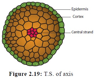
Internal
structure
T.S. of axis
The T.S. of axis shows the presence of epidermis, cortex and central cylinder. The epidermis is the outermost layer and contain chloroplast bearing cells. The cortex is made up of parenchymatous tissue. The cells of the young axis bear chloroplasts. In mature stems the outermost cells become reddish brown colour and become thick walled. Small leaf traces are also noticed. The central cylinder is made up of long, narrow, thin walled, colourless cells which lack protoplasts. They help in the conduction of water and minerals.
T.S. of leaf
A well defined midrib is present. It consists of
several layers of cells but the lateral ‘wing’ or lamina is made up of single
layer of thin walled cells which are rich in chloroplasts. Midrib contains
small strands of slightly thickened narrow cells which help in conduction.
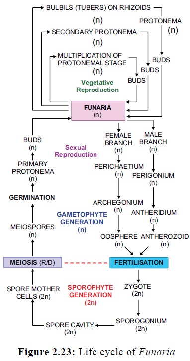
Reproduction
Funaria reproduces
by vegetative and sexual methods.
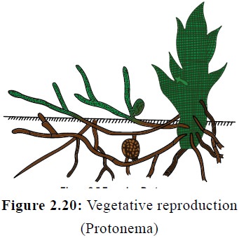
Vegetative reproduction
Vegetative reproduction takes place by the
following methods (Figure 2.20):
1.
Fragmentation of primary protonema,
2.
Formation of secondary protonema from any part of
the gametophyte
3.
Formation of gemmae on terminal cells of the
protonema.
4. Development of Bulbils on the rhizoids.
Sexual reproduction
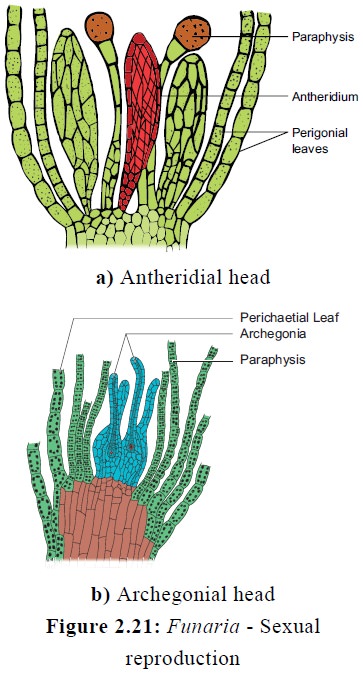
The female sex organ are the archegonia and are borne in clusters on the archegonial branch. Archegonial branches arise laterally at the base of the male branch. They are surrounded by perichaetial leaves. Paraphyses are also present. Each archegonium is flask shaped and is distinguished into a large venter and long neck region. The venter contains venter canal cell and egg. The neck contain neck canal cells (Figure 2.21). Water is essential for fertilization. Rain drops help in the transfer of antherozoids from antheridial head to archegonial head. The antherozoids are attracted to the archegonium through chemotaxis. A large number of antherozoids enter the neck of the archegonium but only one fuses with the egg to form a diploid zygote. The diploid zygote represents the first cell of sporophytic generation and divides to form a sporophyte.
Structure of Sporophyte or capsule
The structure of mature sporophyte of Funaria is complex. The sporophyte is differentiated into foot, seta and capsule (Figure 2.22) . The foot is small, conical and is embedded in the gametophyte.
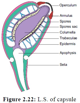
The seta is long, slender and conducts water and nutrients
to the capsule . The capsule is differentiated into apophysis, theca proper and
operculum. The cells constituting the wall of the capsule contain chloroplasts
in them. The apophysis is the lowermost sterile region and connects the capsule
with seta. The epidermis contains stomata which help in exchange of gases. The
cells of the apophysis are photosynthetic, hence the sporophyte of Funaria partially depends on the
gametophyte. The theca proper is the middle part and is fertile region of the
capsule. It consists of a central columella surrounded by spore sac. The spore
sac is surrounded by a single cylindrical air sac traversed by delicate
filaments made up of parenchyma cells called trabeculae. The trabeculae extend from the outer wall of the spore
sac to the innermost layer of the capsule wall. The spore sac contains spore
mother cells which undergo meiotic division to produce haploid spores. The
apical region consists of the operculum and peristome. The operculum is the lid
of the capsule and comes out as a circular cup shaped lid after the dehiscence
of the capsule. The peristome has one or two rows of thickened, tooth like
projections found on the top of the capsule. They are hygroscopic and help in
the dispersal of the spores. During favourble conditions the spores germinate to
produce thread like green branched structure called protonema. It produces
rhizoids and number of lateral buds which develop into new plants. In the life
cycle of Funaria the haploid
gametophytic phase (n)alternates with diploid sporophytic phase (2n) and shows
alternation of generation (Figure 2.23).
Related Topics